Download
1 / 22
270 likes | 512 Views
Imaging Microbubbles. Antony Hsu Shanti Bansal Daniel Handwerker Richard Lee Cory Piette. Topics of Discussion. Brownian Motion What are these bubbles and why do we use them? Following the Great Perrin - Diffusion and Gravitational Motion of Microbubbles Optical Imaging of Microbubbles.

E N D
Imaging Microbubbles Antony Hsu Shanti Bansal Daniel Handwerker Richard Lee Cory Piette
Topics of Discussion • Brownian Motion • What are these bubbles and why do we use them? • Following the Great Perrin - Diffusion and Gravitational Motion of Microbubbles • Optical Imaging of Microbubbles
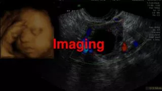
What is ultrasound? • Ultrasound uses high frequency sound waves to image internal structures • The wave reflect off different density liquids and tissues at different rates and magnitudes • It is harmless, but not very accurate
Ultrasound and Microbubbles • Air in microbubbles in the blood stream have almost 0 density and have a distinct reflection in ultrasound • The bubbles must be able to fit through all capillaries and remain stable • We must examine the properties of microbubbles before using this technique
What is Brownian Motion? • Small particles are effected by so many different factors in a solution that they move around at in a random walk • Even if a solution seems stagnate, the microbubbles will still move
What is a Random Walk? • After every seconds, a particle moves in a direction at a velocity v • There is an equal probability that the particle will move in any direction no matter what its past direction was • Each particle is independent of all other particles
Characteristics of Random Walks • Particles have a net displacement of 0 (after time) • Particles usually remain in one region and then wander to other regions
We’re all about Microbubbles (1) Shell Air or High Molecular Weight Gases 1-7mm
We’re all about Microbubbles (4) • Used with ultrasound echocardiography and magnetic resonance imaging (MRI) • Diagnostic imaging - Traces blood flow and outlines images • Drug Delivery and Cancer Therapy
We’re all about Microbubbles (2) Left Arrow: Lipid-Coated Microbubble Right Arrow: Saline Microbubble
Small (1-7 mm) bubbles of air (CO2, Helium) or high molecular weight gases (perfluorocarbon). Enveloped by a shell (proteins, fatty acid esters). Exist - For a limited time only! 4 minutes-24 hours; gases diffuse into liquid medium after use. Size varies according to Ideal Gas Law (PV=nRT) and thickness of shell. We’re all about Microbubbles (5)
Given a volume filled with different sizes of microbubbles, which bubbles move toward which end due to gravity? Following Perrin, we look at the characteristic length (lambda) which will tell us about the motion of the bubble. D l F G = -c(x,t) How Bubbles Separate FT = FD + FG
How do we get lambda(l)? k T meff g rp = density of particle rw = density of water(1g/cm3) r = radius of bubble(cm) meff = (4/3) p r3 (rp - rw) How Bubbles Separate(2) K =Boltzman’s constant (1.38x10⁻²³ J/K) T = Temperature in Kelvin (300K) g = gravity(9.81 m/s2) meff = effective mass l = The size of of microbubbles is known(1-7mm). Therefore, the only factor to be determined is the density of the microbubble. With gas-filled bubbles, the thickness and density of the shell gives the bubble its mass.
How Bubbles Separate(3) • Why is all this important? Well, we want a bubble that will not “float” or “sink.” By adjusting the shell thickness to the force of gravity, we can achieve “neutral buoyancy.” • Basically, by designing the bubble such that the density as a whole has the density of water, then the bubble will undergo only diffusion flux.
Perrin did research on diffusion and brownian motion He conducted experiments to examine diffusion through emulsions He built used a light microscope to visualize emulsions at different depths Perrin determined depth of pictures by the following formula: H=CH’. C = relative refractive index of the two media which the cover-glass separates. H’ = height of microscope. Perrin’s light microscope
Optical target trackingon image sequences • Computer equipment improvement has lead to higher resolution optical imaging • Most computerized optical pattern recognition filters today have been designed to process one image at a time. (isolated images) • These filters would prove ineffective in recording microbubbles moving through the blood stream (image sequencing). • Isolated images do not deal with changes in background, sequential imaging does • this problem leads to the development of the “two image system”--a model that takes into account two successive frames • this model is based on the maximum-likelihood (ML) estimation • The ML estimation takes into account the continuity between two successive frames
Optical target tracking (cont.) • One frame is taken at a known location, one at an “estimated” location • This estimated location will depend on location and size of the object • In this case, the size of microbubble will remain constant (approximately the size of a red blood cell). However, the location will vary. • Idle time between frames depends directly on probability factors. • The two frames are correlated, forming a clear and concise picture of the object’s movement.
A Novel Technique to Visualize Microbubbles • An optical tracking system is placed on Perrin’s light microscope • Allow easy visualization of microbubbles and analysis
Future of Microbubbles • Using microbubbles as a pressure sensitive gauge (especially important for heart) • Enhancing ultrasound/ MR images. Novel gasses used for microbubbles. • Drug delivery