Download
1 / 10
110 likes | 354 Views
ACTINOMYCOSIS Prof. Khaled H. Abu-Elteen. ACTINOMYCETES.

E N D
ACTINOMYCOSIS Prof. Khaled H. Abu-Elteen
ACTINOMYCETES • In this section, we shall discuss 3 genera of actinomycetes: Actinomyces, Nocardia, and Streptomyces. These organisms have been shown to be higher bacteria, but they were thought to be fungi for many years because they havefilamentous forms, 0.5 to 0.8 µ in diameter, which appear to branch. Some species form aerial mycelia in culture. The clinical manifestations of infection are similar to those of a systemic fungal infection. It is now clear that they are not fungi but are closely related to themycobacteria.
Some facts that you should know about these genera are that: • 1- Actinomyces are anaerobic, while Nocardia and Streptomyces are aerobic. • 2- Nocardia stain partially acid-fast, Actinomyces and Streptomyces are not acid-fast. • 3- Actinomyces produce granules. Most actinomycetes in tissue do not stain with the H &E stain commonly used for general histopathology. All genera may produce granules, Actinomyces almost always produce granules.
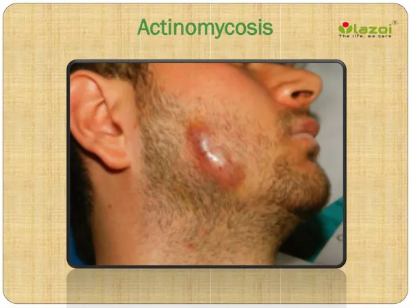
ACTINOMYCOSIS • The most common cause of actinomycosis is the organism Actinomyces israelii which infects both man and animals. In cattle, the disease is called "lumpy jaw" because of the huge abscess formed in the angle of the jaw. In man, A. israelii is an endogenous organism that can be isolated from the mouths of healthy people. Frequently, the infected patient has a tooth abscess or a tooth extraction and the endogenous organism becomes established in the traumatized tissue and causes a suppurative infection.
These abscesses are not confined to the jaw and may also be found in the thoracic area and abdomen. The patient usually presents with a pus-draining lesion, so the pus will be the clinical material you send to the laboratory. This diagnosis can be made on the hospital floor. If you rotate the vial of pus, theyellow sulfur granules, characteristic of this organism, can be seen with the naked eye. You can also see these granules by running sterile water over the gauze used to cover the lesion.
The water washes away the purulent material leaving the golden granules on the gauze. This organism, which occurs worldwide, can be seen histologically as "sulfur granules" surrounded by polymorphonuclear cells (PMN) forming the purulent tissue reaction. The organism is a gram positive rod that frequently branches. The laboratory must specifically be instructed to culture for this anaerobic organism. These lesions must be surgically drained prior to antibiotic therapy and the drug of choice is large doses of penicillin (2 million units q 6 h).
NOCARDIOSIS The most common species of Nocardia which cause disease in human beings are N. brasiliensis and N. asteroides. These are soil organisms which can also be found endogenously in the sputum of apparently healthy people. Nocardiosis primarily presents as a pulmonary disease in the U.S. In Latin America, it is more frequently seen as the cause of a subcutaneous infection, with or without draining abscesses. It can even present as a lesion in the chest wall that drains onto the surface of the body similar to actinomycosis. Brain abscesses are frequent secondary lesions. N.asteroides is usually the etiologic agent of pulmonary nocardiosis while N. brasiliensis is frequently the cause of sub-cutaneous lesions.
The material sent to the lab,depending on the presentation of the disease, is sputum, pus, or biopsy material. • These organismsrarely form granules. TheNocardia areaerobic, gram-positive rodsand stainpartially acid-fast (i.e., the acid-fast staining is not uniform). Thereare no serological tests, and thedrug of choice is Sulfa drugs (Trimethoprim). The nocardia grow readily on most bacteriologic and TB media. The geographic distribution of these organisms is worldwide.
STREPTOMYCETES The streptomyces species usually cause the disease entity known as mycetoma (fungus tumor). These infections are usually subcutaneous, but they can penetrate deeper and invade the bone. Some species produce a protease which inhibits macrophages. Material sent to the lab is pus or skin biopsy. The streptomycetes are aerobic like Nocardia, and can grow on both bacterial and fungal (SDA) media. They produce a chalky aerial mycelium with much branching. It is important to let the lab know the organism you suspect because most bacterial pathogens will grow out overnight, but the actinomycetes take longer to be visible on the culture plates (48-72 h).
The various species of streptomyces produce granules of different size, texture and color. These granules along with colonial growth and biochemical tests allow the bacteriologist or mycologist to identify each species. The organisms are found world-wide. There are no serological tests, and the drugs of choice are the • combination of sulfamethoxazole/trimethoprim or amphotericin B. In the tropics this disease may go undiagnosed or untreated for so long that surgical amputation may be the only effective treatment.