Download
1 / 81
830 likes | 1.48k Views
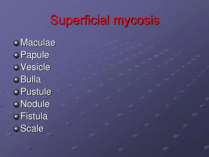
Superficial mycosis. Maculae Papule Vesicle Bulla Pustule Nodule Fistula Scale. Superficial mycosis. Definition Fungi Pityriasis versicolor Tinea nigra Piedra Bacteria Erythrasma Trichomycosis axillaris. Superficial mycosis. Otomycosis Keratomycosis Pitted keratolysis

E N D
Superficial mycosis • Maculae • Papule • Vesicle • Bulla • Pustule • Nodule • Fistula • Scale
Superficial mycosis • Definition • Fungi • Pityriasis versicolor • Tinea nigra • Piedra • Bacteria • Erythrasma • Trichomycosis axillaris
Superficial mycosis • Otomycosis • Keratomycosis • Pitted keratolysis • Dermatophylosis
Pityriasis versicolor • Tinea versicolor • Maculae • Malassezia spp • Normal flora • Boys & girls • After maturation • Beauty • Warm & humidity • Season
Pityriasis versicolor • Conditions: • Health • Sweat • Greasy skin • IC. • Chronic bacterial infections • Steroids
Pityriasis versicolor • Clinical manifestations: • Maculae • White, cream, pink, red, brown • Position • Scale • Painless • Not itching
Superficial mycosis • Pityriasis versicolor
Superficial mycosis • Pityriasis versicolor
Superficial mycosis • Pityriasis versicolor
Superficial mycosis • Pityriasis versicolor
Superficial mycosis • Pityriasis versicolor
Superficial mycosis • Pityriasis versicolor
Superficial mycosis • Pityriasis versicolor
Superficial mycosis • Pityriasis versicolor
Superficial mycosis • Pityriasis versicolor
Pityriasis versicolor • Differential diagnosis: • Vitiligo • Chloasma
Pityriasis versicolor • Laboratory diagnosis: • Sampling • Scalpel • Scathe tape • Wood ́s lamp • Direct • Culture
Pityriasis versicolor • Treatment • Selenium sulfide • Clotrimazole
Tinea nigra • Phaeoanellomyces werneckii • Exophiala werneckii • Ecology • Palm • Tinea nigra palmaris
Tinea nigra • Clinical manifestation: • Annular • Centrifuge • Regular or irregular • Painless • No itching
Tinea nigra • Differential diagnosis: • Malignant melanoma • Silver nitrate
Tinea nigra • Laboratory diagnosis: • Sampling: • Scalpel & KOH10% • Direct: • Hyphae • Chlamydioconidia • Culture: • S
Tinea nigra • Treatment • Keratolytic ointment • whitfield
Piedra • Hair • Nodule • Bread, groin, axillar • White & black
Piedra • Clinical manifestation: • White piedra: • Trichosporon beigelii • Basidiomycetes • Nodule • Bread, head, pubis • White, brown, yellow, • Soft
Piedra • Clinical manifestation: • Black piedra: • Piedra hortae • Ascomycetes • Nodule • Head • Thick • Black to brown
Piedra • Differential diagnosis: • Pediculosis
Piedra • Laboratory diagnosis: • Sampling: • Sesser & KOH10% • Direct: • White: hyphae & arthroconidia • Black: ascus & ascospore • Culture: • S
Piedra • Treatment: • Scraping • Clotrimazole
Erythrasma • Bacteria • Corynebacterium minutissimum • Chronic • Intertrigenous
Erythrasma • Clinical manifestation: • Maculae • Red or brown • Scale • Itching • No inflammation
Superficial mycosis Erythrasma
Superficial mycosis Erythrasma
Superficial mycosis Erythrasma
Superficial mycosis Erythrasma
Erythrasma • Differential diagnosis: • Pityriasis versicolor • Candidiasis • Tinea cruris
Erythrasma • Laboratory diagnosis: • Sampling: • Scalpel & simple staining • Direct: • Strand • Culture: • BHI, Blood agar • Wood ́s lamp
Erythrasma • Treatment • Erythromycin
Trichomycosis axillaris • Bacteria • Corynebacterium tenuis • Nodule • Hair • Axillaries & pubis • Red • Yellow • black
Trichomycosis axillaris • Differential diagnosis: • Pediculosis
Trichomycosis axillaris • Laboratory diagnosis: • Sampling: • Sesser & KOH10% • Direct • Culture
Trichomycosis axillaris • Treatment • Scraping and sulphur ointment 3%
Keratomycosis FUNGAL KERATITIS (FK)
INCIDENCE OF FK • Developed world 6 -35% of all microbial keratitis • Developing world 22 - >50%
FUNGAL GROUPS • Filamentous Fusarium Aspergillus Dematiaceous • Yeasts Candida
Fungal Keratitis • Risk factors: • Topical corticosteroids ↓corneal resistance to infection • Contact lens use • Immunocomprised states
Fungal Keratitis • Risk factors • Injury, ocular surface compromise • Temperature • Wind • Humidity • Urbanisation/employment
Fungal Keratitis • Exogen • Endogen
Fungal Keratitis • Gray-white color, dry, and rough corneal surface • White ring in the cornea and satellite lesions near the edge of the primary • Most cases remain superficial but deep invasion may occur