Download
1 / 118
1.19k likes | 1.78k Views
Introduction. Otosclerosis Primary metabolic bone disease of the otic capsule and ossiclesResults in fixation of the ossicles and conductive hearing lossMay have sensorineural component if the cochlea is involvedGenetically mediatedAutosomal dominant with incomplete penetrance (40%) and variab

E N D
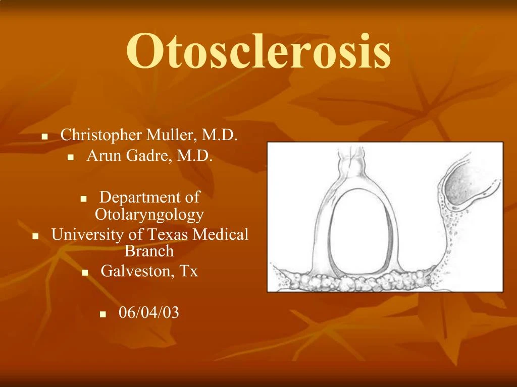
1. Otosclerosis
Christopher Muller, M.D.
Arun Gadre, M.D.
Department of Otolaryngology
University of Texas Medical Branch
Galveston, Tx
06/04/03
2. Introduction Otosclerosis
Primary metabolic bone disease of the otic capsule and ossicles
Results in fixation of the ossicles and conductive hearing loss
May have sensorineural component if the cochlea is involved
Genetically mediated
Autosomal dominant with incomplete penetrance (40%) and variable expressivity
3. History of Otosclerosis and Stapes Surgery 1704 � Valsalva first described stapes fixation
1857 � Toynbee linked stapes fixation to
hearing loss
1890 � Katz was first to find microscopic
evidence of otosclerosis
1893 � Politzer described the clinical entity of
�otosclerosis�
4. History of Otosclerosis and Stapes Surgery Gunnar Holmgren
Father of fenestration surgery
Single stage technique
Sourdille
Holmgren�s student
3 stage procedure
64% satisfactory results
5. History of Otosclerosis and Stapes Surgery Julius Lempert
Popularized the single staged fenestration procedure
John House
Further refined the procedure
Popularized blue lining the horizontal canal
6. History of Otosclerosis and Stapes Surgery
Fenestration procedure for otosclerosis
Fenestration in the horizontal canal with a tissue graft covering
>2% profound SNHL
Rarely complete closure of the ABG
7. History of Otosclerosis and Stapes Surgery Samuel Rosen
1953 � first suggest mobilization of the stapes
Immediate improved hearing
Re-fixation
8. History of Otosclerosis and Stapes Surgery John Shea
1956 � first to perform stapedectomy
Oval window vein graft
Nylon prosthesis from incus to oval window
9. Epidemiology
10% overall prevalence of histologic otosclerosis
1% overall prevalence of clinically significant otosclerosis
10. Epidemiology % incidence of
Race otosclerosis
Caucasian 10%
Asian 5%
African American 1%
Native American 0%
11. Epidemiology Gender
Histologic otosclerosis � 1:1 ratio
Clinical otosclerosis � 2:1 (W:M)
Increase progression during pregnancy (10%-17%)
Bilaterality more common (89% vs. 65%)
12. Epidemiology Age
15-45 most common age range of presentation
Youngest presentation7 years
Oldest presentation 50s
0.6% of individuals <5 years old have foci of otosclerosis
13. Pathophysiology
Osseous dyscrasia
Resorption and formation of new bone
Limited to the temporal bone and ossicles
Inciting event unknown
Hereditary, endocrine, metabolic, infectious, vascular, autoimmune, hormonal
14. Pathophysiology Siebenmann � first to describe the microscopic appearance
Spongy
Usually limited to the anterior footplate
15. Pathology Two phases of disease
Active (otospongiosis phase)
Osteocytes, histiocytes, osteoblasts
Active resorption of bone
Dilation of vessels
Schwartze�s sign
Mature (sclerotic phase)
Deposition of new bone (sclerotic and less dense than normal bone)
18. �Blue mantles of Manasseh�
19. Pathophysiology
20. Pathology
Most common sites of involvement
Fissula ante fenestrum
Round window niche (30%-50% of cases)
Anterior wall of the IAC
21. Fissula ante and post fenestrum
22. Fissula ante fenestrum
23. Non-clinical foci of otosclerosis
24. Annular ligament involvement
25. Footplate Involvement
26. Anterior footplate involvement
27. Bipolar involvement of the footplate
29. Round Window
30. Labyrinthine Otosclerosis 1912 � Siebenmann described labyrinthine otosclerosis
Suggested otosclerosis may cause SNHL
Toxic metabolites
Decreased blood supply
Direct extension
31. IAC
32. Hyalinization of the spiral ligament
33. Erosion into inner ear
36. Organ of Corti
37. Diagnosis
38. History Most common presentation
Women in her 20s or 30s
Conductive or Mixed hearing loss
Slowly progressive,
Bilateral (80%)
asymmetric
Tinnitus (75%)
39. History Age of onset of hearing loss
Progression
Laterality
Associated symptoms
Dizziness
Otalgia
Otorrhea
Tinnitus
40. History
Vestibular symptoms
25%
Most commonly dysequilibrium
Occasionally attacks of vertigo with rotatory nystagmus
Prior otologic surgery
History of ear infections
41. History Family history
2/3 have a significant family history
Particularly helpful in patients with severe or profound mixed hearing loss
42. Physical Exam Otomicroscopy
Most helpful in ruling out other disorders
Middle ear effusions
Tympanosclerosis
Tympanic membrane perforations
Cholesteatoma or retraction pockets
Schwartze�s sign
Red hue in oval window niche area
10% of cases
Pneumatic otoscopy
Distinguish from malleus fixation
43. Physical Exam Tuning forks
Hearing loss progresses form low frequencies to high frequencies
256, 512, and 1024 Hz TF should be used
Rinne
256 Hz � negative test indicates at least a 20 dB ABG
512 Hz � negative test indicates at least a 25 dB ABG
44. Differential Diagnosis Ossicular discontinuity
Congenital stapes fixation
Malleus head fixation
Paget�s disease
Osteogenesis imperfecta
45. Audiometry Tympanometry
Impedance testing
Acoustic reflexes
Pure tones
46. Tympanometry Jerger (1970) � classification of tympanograms
Type A
Type A
Type As
Type Ad
Type B
Type C
48. Acoustic Reflexes Result from a change in the middle ear compliance in response to a sound stimulus
Change in compliance
Stapedius muscle contraction
Stiffening of the ossicular chain
Reduces the sound transmission to the vestibule
49. Acoustic Reflexes Otosclerosis has a predictable pattern of abnormal reflexes over time
Diphasic reflex pattern
Reduced reflex amplitude
Elevation of ipsilateral thresholds
Elevation of contralateral thresholds
Absence of reflexes
50. Acoustic Reflexes
51. Pure Tone Audiometry
Most useful audiometric test for otosclerosis
Characterizes the severity of disease
Frequency specific
52. Pure Tone Audiometry Low frequencies affected first
Below 1000 Hz
Rising air line
�Stiffness tilt�
Secondary to stapes fixation
53. Pure Tone Audiometry With disease progression
Air line flattens
Secondary to mass effect
55. Pure Tone Audiometry Carhart�s notch
Hallmark audiologic sign of otosclerosis
Decrease in bone conduction thresholds
5 dB at 500 Hz
10 dB at 1000 Hz
15 dB at 2000 Hz
5 dB at 4000 Hz
56. Pure Tone Audiometry Carhart�s notch
Proposed theory
Stapes fixation disrupts the normal ossicular resonance (2000 Hz)
Normal compressional mode of bone conduction is disturbed because of relative perilymph immobility
Mechanical artifact
Reverses with stapes mobilization
57. Imaging Computed tomography (CT) of the temporal bone
Proponents of CT for evaluation of otosclerosis
Pre-op
Characterize the extent of otosclerosis
Severe or profound mixed hearing loss
Evaluate for enlarge cochlear aqueduct
Post-op
Recurrent CHL
Re-obliteration vs. prosthesis dislocation
Vertigo
58. Imaging CT
Axial cuts
Patient position � canthomeatal line perpendicular to the table top
1 mm cuts
Top of sup. SCC to bottom of the cochlea
Coronal
Patient position � supine w/ head overextended
face turned 20 degrees ipsilateral
60. �Halo sign�
61. Paget�s disease
62. Osteogenesis Imperfecta
63. Natural history of otosclerosis 90% of all cases are never clinically apparent
Foci begins in childhood
Most commonly becomes symptomatic in the 3rd and 4th decades
After clinical presentation
Conductive hearing loss progressive
Periods of quiescence and deterioration
Worsening tinnitus
Associated SNHL (rarely purely SN)
Matures by age 50-70 with max. CHL of 50 dB
64. Management Medical � Sodium Fluoride
Amplification
Surgery
Combinations
65. Patient Selection Factors
Result of TF tests and audiometry
Skill of the surgeon
Facilities
Medical condition of the patient
Patient wishes
66. Patient Counseling Options for treatment
Advantages and disadvantages of each
Repeat clinic visit
67. Surgery Best surgical candidate
Previously un-operated ear
Good health
Unacceptable ABG
25 to 40 dB, bilateral ABG recommended by different authorities
Negative Rinne test
Excellent discrimination
Desire for surgery
68. Surgery Other factors
Age of the patient
Elderly
Poorer results in the high frequencies
Congenital stapes fixation (44% success rate)
Juvenile otosclerosis (82% success rate)
Occupation
Diver
Pilot
Airline steward/stewardess
69. Surgery Other factors
Vestibular symptoms
Meniere's disease
Concomitant otologic disease
Cholesteatoma
Tympanic membrane perforation
70. Endolymphatic Hydrops
71. Surgical Steps Subtleties of technique and style
Local vs. general anesthesia
Stapedectomy vs. partial stapedectomy vs. stapedotomy
Laser vs. drill vs. cold instrumentation
Oval window seals
Prosthesis
72. Pre-op
Confirm the correct ear (largest ABG)
With the patient
Audiogram
History and physical exam
Place CT and audiogram in a visible location in the OR for easy intra-operative evaluation
73. Canal Injection 2-3 cc of 1% lidocaine with 1:50,000 or 1:100,000 epinephrine
4 quadrants
Bony cartilaginous junction
74. Raise Tympanomeatal Flap 6 and 12 o�clock positions
6-8 mm lateral to the annulus
Take into account curettage of the scutum
75. Separation of chorda tympani nerve from malleus Separate the chorda from the medial surface of the malleus to gain slack
Avoid stretching the n.
Cut the nerve rather than stretch it
76. Curettage of Scutum Curettage a trough lateral to the scutum, thinning it
Then remove the scutum (incus to the round window)
Visualize the pyramidal process and facial n.
77. Curettage of Scutum Exposure of pyramidal process and facial n.
Preservation of bone over incus
78. Middle ear examination Mobility of ossicles
Confirm stapes fixation
Evaluate for malleus or incus fixation
Abnormal anatomy
Dehiscent facial nerve
Overhanging facial nerve
Deep narrow oval window niche
79. Measurement for prosthesis Measurement
Lateral aspect of the long process of the incus to the footplate
Average 4.5 mm
81. Total Stapedectomy Uses
Extensive fixation of the footplate
Floating footplate
Disadvantages
Increased post-op vestibular symptoms
More technically difficult
Increased potential for prosthesis migration
86. Stapedotomy/Small Fenestra Originally for obliterated or solid footplates
Europe
1970-80
First laser stapedotomy performed by Perkins in 1978
Advantages
Less trauma to the vestibule
Less incidence of prosthesis migration
Less fixation of prosthesis by scar tissue
87. Drill Fenestration 0.7mm diamond burr
Motion of the burr removes bone dust
Avoids smoke production
Avoids surrounding heat production
88. Laser Fenestration Laser
Avoids manipulation of the footplate
Argon and Potassium titanyl phosphate (KTP/532)
Wave length 500 nm
Visible light
Absorbed by hemoglobin
Surgical and aiming beam
Carbon dioxide (CO2)
10,000 nm
Not in visible light range
Surgical beam only
Requires separate laser for an aiming beam (red helium-neon)
Ill defined fuzzy beam
91. Fenestration Causse et al. (1993)
Recommends posteriorly placed fenestration to better recreate the natural physiologic dynamics of the footplate
92. Pivoting stapes
93. Energy transmission to the stapes
94. Posterior Fenestration Posteriorly placed fenestration with the laser
Causse also recommends following the laser with the diamond burr to remove char
95. Oval window seal Tragal perichondrium
Vein (hand or wrist)
Temporalis fascia
Blood
Fat
96. Vein graft
97. Reconstructing the annular ligament
98. Placement of the Prosthesis Prosthesis is chosen and length picked
Some prefer bucket handle to incorporate the lenticular process of the incus
99. Stapedectomy vs. Stapedotomy ABG closure < 10dB (PTA)
100. Stapedectomy vs. Stapedotomy ABG closure at 4 kHz
101. Special Considerations and Complications in Stapes Surgery
102. Overhanging Facial Nerve Usually dehiscent
Consider aborting the procedure
Facial nerve displacement (Perkins, 2001)
Facial nerve is compressed superiorly with No. 24 suction (5 second periods)
10-15 sec delay between compressions
Perkins describes laser stapedotomy while nerve is compressed
Wire piston used
Add 0.5 to 0.75 mm to accommodate curve around the nerve
103. Floating Footplate Footplate dislodges from the surrounding OW niche
Incidental finding
More commonly iatrogenic
Prevention
Laser
Footplate control hole
Management
Abort
H. House favors promontory fenestration and total stapedectomy
Perkins favors laser fenestration
104. Floating Footplate Hearing results
Thin or blue footplate � 97% ABG closure (<10dB)
White or �biscuit� footplate � 52% ABG closure
105. Diffuse Obliterative Otosclerosis Occurs when the footplate, annular ligament, and oval window niche are involved
Bone is thinned with a small cutting burr
Blue lined at anteroposterior edges first
106. Perilymphatic Gusher Associated with patent cochlear aqueduct
More common on the left
Increased incidence with congenital stapes fixation
Increases risk of SNHL
Management
Ruff up the footplate
Rapid placement of the OW seal then the prosthesis
HOB elevated, stool softeners, bed rest, avoid Valsalva, +/- lumbar drain
107. Round Window Closure 20%-50% of cases
1% completely closed
No effect on hearing unless 100% closed
Opening has a high rate of SNHL
108. SNHL 1%-3% incidence of profound permanent SNHL
Surgeon experience
Extent of disease
Cochlear
Prior stapes surgery
Temporary
Serous labyrinthitis
Reparative granuloma
Permanent
Suppurative labyrinthitis
Extensive drilling
Basilar membrane breaks
Vascular compromise
Sudden drop in perilymph pressure
109. Reparative Granuloma Granuloma formation around the prosthesis and incus
2 -3 weeks postop
Initial good hearing results followed by an increase in the high frequency bone line thresholds
Associated tinnitus and vertigo
Exam � reddish discoloration of the posterior TM
Treatment
ME exploration
Removal of granuloma
Prognosis � return of hearing with early excision
110. Vertigo Most commonly short lived (2-3 days)
More prolonged after stapedectomy compared to stapedotomy
Due to serous labyrinthitis
Medialization of the prosthesis into the vestibule
With or without perilymphatic fistula
Reparative granuloma
112. Recurrent Conductive Hearing Loss Slippage or displacement of the prosthesis
Most common cause of failure
Immediate
Technique
Trauma
Delayed
Slippage from incus narrowing or erosion
Adherence to edge of OW niche
Stapes re-fixation
Progression of disease with re-obliteration of OW
Malleus or incus ankylosis
115. Amplification Amplification
Excellent alternative
Non-surgical candidates
Patients who do not desire surgery
Satisfaction rate less than with successful Sx
Canal occlusion effect
Amplification not used at night
116. Medical Sodium Fluoride
1923 - Escot suggested using calcium fluoride
1965 � Shambaugh popularized its use
Mechanism
Fluoride ion replaces hydroxyl group in bone forming fluorapatite
resistant to resorption
Increases calcification of new bone
Causes maturation of active foci of otosclerosis
117. Sodium Fluoride
Reduces tinnitus, reverses Schwartze�s sign, resolution of otospongiosis seen on CT
OTC � Florical
Dose � 20-120mg
Indications
Non-surgical candidates
Patients who do not want surgery
Surgical candidates with + Schwartze�s sign
Treat for 6 mo pre-op
Postop if otospongiosis detected intra-op
118. Sodium fluoride
Hearing results
50% stabilize
30% improve
Re-evaluate q 2 yrs with CT and for Schwartze�s sign to resolve
If fluoride are stopped � expect re-activation within 2-3 years
119. References Bibliography
Causse JR et al. Sodium fluoride therapy. Am J Otol 1993;14(5):482-490
Glasscock II ME, et al. Twenty-five years of experience with stapedectomy.
Laryngoscope 1995;105:899-904
House HP, Kwartler JA. Total stapedectomy. Otologic Surgery, 2nd ed. edited by
Brackmann, Shelton, and Arriaga, W.B. Saunders 2001;226-234
Hough J. Partial stapedectomy. Ann Otol Rhinol Laryngol 1960;69:571
House J. Otosclerosis. Otolaryngol Clinics 1993;26(3):323-502
Jerger J. Clinical experience with impedance audiometry. Arch Otolaryngol
1970;92:311
Lempert J. Improvement in hearing in cases of otosclerosis: A new, one stage surgical
technique. Arch Otolaryngol 1938;28:42-97
Lippy WH, Schuring AG. Treatment of the inadvertently mobilized footplate.
Otolaryngol Head Neck Surg 1973;98:80-81
Meyer S. The effect of stapes surgery on high frequency hearing in patients with
otosclerosis Am J Otol 1999;20:36-40
Millman B. Giddings, NA and Cole, JM. Long-term follow-up stapedectomy in children
and adolescents. Otol Head Neck Surg 1996;115(1):78-81
Perkins RC. Laser stapedotomy. Otologic Surgery, 2nd ed. edited by Brackmann,
Shelton, and Arriaga, W.B Saunders 2001;245-260
Perkins RC. Laser stapedotomy for otosclerosis. Laryngoscope 1980;91:228-241
Roland PS. Otosclerosis. www.emedicine.com/ped/topic1692.htm. 2002;1-11
Roland PS, Meyerhoff WL. Otosclerosis. Otolaryngology-Head and Neck Surgery. 3rd
ed., edited by Byron J. Bailey, Lippincott Williams & Wilkins, Philadelphia
2001;1829-1841
Rosen S. Restoration of hearing in otosclerosis by mobilization of the fixed stapedial
footplate. An analysis of results. Laryngoscope 1955;65:224-269
Shea J Jr. Fenestration of the oval window. Ann Otol Rhinol Laryngol 1958;67:932-951
Shambaugh G. Clinical diagnosis of cochlear (labyrinthine) otosclerosis. Laryngoscope
1965;75:1558-1562
Shambaugh GE, Jr. and Glasscock ME, III. Surgery of the ear, 3rd ed. Philadelphia, W.
B. Saunders, 1980;455-516