Download
1 / 27
270 likes | 353 Views
GRID REQUIREMENTS OF IN SILICO ONCOLOGY: THE ACGT PROJECT PARADIGM G. Stamatakos gestam@central.ntua.gr. EGEE’08 CONFERENCE 22-26 Sept. 2008. ACKNOWLEDGMENTS. Special thanks are due to Dimitra Dionysiou, ICCS-National Technical University of Athens Manolis Tsiknakis, FORTH, Greece

E N D
GRID REQUIREMENTS OF IN SILICO ONCOLOGY: THE ACGT PROJECT PARADIGMG. Stamatakosgestam@central.ntua.gr EGEE’08 CONFERENCE 22-26 Sept. 2008
ACKNOWLEDGMENTS Special thanks are due to • Dimitra Dionysiou, ICCS-National Technical University of Athens • Manolis Tsiknakis, FORTH, Greece • Norbert Graf, Dept. Hematology and Oncology, University of Saarland • and • the whole ACGT Consortium for their multifaceted contribution to the development of the Oncosimulator and its positioning within the overall ACGT architecture
3rd International Advanced Research Workshop on In Silico Oncology: Advances and Challenges • September 23- 24, 2008 • Zografeio Lyceum, Istanbul, Turkey • http://www.3rd-iarwiso.iccs.ntua.gr/
WHAT IS THE ACGT“ONCOSIMULATOR” AND WHAT IS ITS PURPOSE ? • The “ACGT ONCOSIMULATOR” is a a • clinically meaningful, • scientifically sound, • technologically advanced and • user friendly system able to spatiotemporally simulate within well defined reliability limits tumour growth and tumour and [to a lesser extent] normal tissue response to (chemo)therapeutic schemes for the cases of breast cancer and nephroblastoma (Wilms’ tumour) in the patient’s individualized context.
The final goal of the the “Oncosimulator” is to contribute to the optimization of cancer therapeutic strategies through conducting in silico (=on the computer) experiments based on the individual patient’s multilevel data in order to support the optimal selection of the treatment scheme to be administered. • An additional target is to support the design and interpretation of new clininicogenomic trials.
PURPOSE OF THE DEMONSTRATOR • To show how the “Oncosimulator” i.e. the ACGT in silico oncology component would look like and function. • To show how real clinical trials can be simulatedin silico through the integration of basic science knowledge. • To outline how Grid services are expected to be utilized by in silico oncology. • To outline how advanced visualization techniques can be used in order to make predictions easily understood in the four dimensional context
As the ACGT medical data [imaging, histopathological, molecular and clinical] are still in the process of reliable, safe, legal and ethical distribution within the ACGT consortium, • the “Oncosimulator” concept and construct is demonstrated using previous work done at ICCS during the last ten years coupled with new work done by ICCS, PSNC, UvΑ and other partners within the framework of ACGT.
CLINICAL CONTEXT OF THE DEMONSTRATOR • Two arms of the Radiation Therapy Oncology Group (RTOG) Clinical Study 83-02 are simulated in silico. • The study concerns glioblastoma multiforme treated with radiotherapy [17] • Imaging data [from the Whole Brain Atlas ] concerning a glioblastoma have been used along with the radiobiological alpha and beta values that correspond to a specific glioblastoma cell line with mutant p53 gene [molecular data] [17] • The exact type of tumour [glioblastoma multiforme] corresponds to the histopathological data here.
THE TWO RTOG STUDY 83-02 SIMULATED • 1) AHF-48Gy: accelerated hyperfractionation, 48Gy total dose, (1.6Gy twice daily to a total dose of 48 Gy) • 2) HF-81.6Gy: hyperfractionation, 81.6Gy total dose.(1.2Gy twice daily to a total dose of 81.6Gy)
The RTOG study 83-02 [1] was a randomized Phase I/II study of escalating doses for Hyperfractionated radiotherapy (HF, 1.2Gy twice daily to doses of 64.8, 72, 76.8, or 81.6Gy) and Accelerated Hyperfractionated radiotherapy (AHF, 1.6Gy twice daily to doses of 48 or 54.4Gy) with carmustine (BCNU) for adults with supratentorial glioblastoma multiforme (GBM) or anaplastic astrocytoma. • The study has revealed that GBM patients who received the higher HF doses had survival superior to the patients in the AHF arms or lower HF doses. • In silico experiments corresponding to the different arms of RTOG 83-02 study have been performed by ISOG/ICCS/NTUA [17].
RADIOBIOLOGICAL DATA BASED ON THE GBM GENETIC PROFILE (FROM EXPERIMENTAL WORK) • In the results presented here, a hypothetical GBM tumour with mutant (mt) p53 gene is considered [2]: αp= 0.17 Gy-1, βP=0.02 Gy-2 • (we also set αG0 = αP /OER, βG0= βP/OER2, OER = 3, and αS = 0.6 αP + 0.4 αG0 , βS= 0.6 βP + 0.4 βG0,[3],[4] (p.99)). • The meaning of the symbols used is the following: • αp, βP: the LQ Model parameters for all proliferative cell cycle phases except for the DNA synthesis phase (S phase). • αS, βS: the LQ Model parameters for the S phase. • αG0, βG0 : the LQ Model parameters for the resting G0 phase.
Typical clonogenic cell densities are 104 to 105 cells/mm3 [5]. • Since most GBM tumours are poorly differentiated and rapidly growing, we assume a clonogenic cell density of 2105 cells/mm3 in the proliferating cell region, 105 cells/mm3 in the G0 cell region and 0.2105 cells/mm3 in the dead cell region of the tumour. • The cell cycle duration has been taken equal to 40h. This is the average of the cell cycle durations we have found in the literature for GBM cell lines [6],[7]. • In [8] the approximate percentage of the cell cycle time spent in each phase by a typical malignant cell is assumed as follows: TG1 = 0.4 TC, TS = 0.39TC, TG2 = 0.19TC, TM = 0.02TC. The duration of the G0 phase is taken to be TG0=25h [9].
The cell loss factor (CLF) is considered equal to 0.3 [10]. In [11] the authors note thatcell loss is mainly due to necrosis (CLFN) and apoptosis (CLFA) and that gliomas have a low CLF in general. • We assume that the total CLF (0.3) is the sum of the CLFN (0.27) and CLFA (0.03). • We hypothesize low levels of apoptotic cells for GBM, as we have found that this is in general the case for gliomas [2],[11],[12]. )
CLINICAL TRIAL SPECIFICS • The delivery of irradiation takes place at 08:00 and 16:00 every day, 5 days per week (no irradiation during weekends). • The distribution of the absorbed dose in the tumour region is assumed to be uniform. • It should also be noted that carmustin, which was administered to all patients enrolled in the RTOG – 83-02 study, is assumed not to significantly modify the relative effectiveness of the radiation therapy schedules considered, as the chemotherapy administration schedule was the same for all patients.
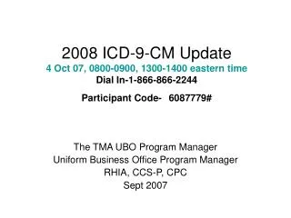
Figure 1 presents the results of the in silico experiments in the form of the number of surviving tumour cells (proliferating and dormant) as a function of time (Last time point: 8 weeks after the beginning of the radiotherapy treatment, t=1344h). • Improved tumour control following high-dose HF irradiation is evident in the diagram and in agreement with the conclusions of the clinical trial. • In fact (data not shown here), the higher the total dose in an HF schedule, the better the result in terms of tumour cell kill.
Figure 1. Number of surviving tumour cells as a function of time for a glioblastoma tumour with mutant p53 gene. AHF-48Gy: accelerated hyperfractionation, 48Gy total dose, HF-81.6Gy: hyperfractionation, 81.6Gy total dose.
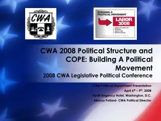
Initial tumour region at the beginning of the radiotherapy treatment
8 weeks later (t=1344h) Irradiation according to HF 81.6Gy 8 weeks later (t=1344h) Irradiation according to AHF 48Gy Colour code → red: proliferating cell region, green: G0 cell region, blue: dead cell region. See [17] for details
More specifically, the inspection of the simulation results reveals that AHF schedules, which employ a higher fraction dose compared to HF schedules, seem at first to be beneficial as they achieve the maximum tumour cell kill at some instant. • Nevertheless, the duration of the AHF schedules is smaller; as a result, if they fail in eradicating “all” tumour cells, tumour repopulation begins earlier.
Descriptions of various aspects of the “top-down” simulation approach developed at the In Silico Oncology Group Institute of Communication and Computer Systems, Natl. Tech.Univ.of Athens [www.in-silico-oncology.iccs.ntua.gr ]and applied on the clinical cases considered can be found in publications [13-16] and in particular in [17].
Need for code execution on Grid environment • Use of Grid architectures is important in order to execute the code for a very large number of parameters values combinations concurrently. • This is necessary in order to increase the reliability of the “Oncosimulator” as many parameter values are not known but only their ranges can be estimated.
REFERENCES • [1] Werner-Wasik M. et al “Final report of a phase I/II trial of hyperfractionated and accelerated hyperfractionated radiation therapy with carmustine for adults with supratentorial malignant gliomas”, Cancer 77, 1535-1543, 1996. • [2] D.A. Haas-Kogan, G. Yount, M. Haas, D. Levi, S.S. Kogan, L. Hu, C. Vidair, D.F. Deen, W.C. Dewey, M.A. Israel, p53-dependent G1 arrest and p53 independent apoptosis influence the radiobiologic response of glioblastoma, Int. J. Radiat. Oncol. Biol. Phys. 36, 95-103, 1996. • [3] M. Kocher, H. Treuer, J. Voges, M. Hoevels, V. Sturm, R.P. Mueller, Computer simulation of cytotoxic and vascular effects of radiosurgery in solid and necrotic brain metastases, Radiother. Oncol. 54, 149-156, 2000. • [4] C. Perez and L. Brady, Principles and Practice of Radiation Oncology. Philadelphia: Lippincott-Raven, 1998. • [5] B. Jones and R.G. Dale, Mathematical models of tumour and normal tissue response, Acta Oncol.38, 883-893, 1999. • [6] L.E. Dillehay, A model of cell killing by low-dose-rate radiation including repair of sublethal damage, G2 block, and cell division, Rad. Res.124, 201-207, 1990.
REFERENCES (cont.) • [7] B. Hegedues, A. Czirok, I. Fazekas, T. Babel, E. Madarasz, T. Viscsek, Locomotion and proliferation of glioblastoma cells in vitro: statistical evaluation of videomicroscopic observations, J. Neurosurgery92, 428-434, 2000. • [8] B.G. Katzung (ed), Basic and Clinical Pharamacology. International Edition. 2001. • [9] W. Duechting, W. Ulmer, R. Lehrig, T. Ginsberg, E. Dedeleit, Computer simulation and modelling of tumour spheroid growth and their relevance for optimization of fractionated radiotherapy, Strahlenther. Onkol. 168, 354-360, 1992. • [10] P. Huang, A. Allam, L. Perez, A. Taghian, J. Freeman, H. Suit, The effect of combining recombinant human tumor necrosis factor-alpha with local radiation on tumor control probability of a human glioblastoma multiforme xenograft in nude mice, Int J Radiat Oncol Biol Phys32(1), 93-98, 1995. • [11] M. Nakajima, S. Nakasu, S. Morikawa, T. Inubushi, Estimation of volume doubling time and cell loss in an experimental rat glioma model in vivo, Acta Neurochir140, 607-613, 1998. • [12] S. Tribius, A. Pidel, D. Casper, ATM protein expression correlates with radioresistance in primary glioblastoma cells in culture,Int. J. Radiat. Oncol. Biol. Phys. 50, 511-523, 2001.
REFERENCES (cont.) • [13] G.S.Stamatakos, D.D.Dionysiou, E.I.Zacharaki, N.A.Mouravliansky, K.Nikita, N.Uzunoglu, “In silico radiation oncology: combining novel simulation algorithms with current visualization techniques”, Proceedings of the IEEE, vol. 90, No11, Nov. 2002. 1764-1777 • [14] D. D. Dionysiou, G. S. Stamatakos, N.K. Uzunoglu, K. S. Nikita, A. Marioli, “A four-dimensional simulation model of tumour response to radiotherapy in vivo: parametric validation considering radiosensitivity, genetic profile and fractionation,” Journal of Theoretical Biology 230 (2004) 1–20 • [15] V. P Antipas, G. S Stamatakos, N. K Uzunoglu, D. D Dionysiou, R. G Dale, ” A spatio-temporal simulation model of the response of solid tumours to radiotherapy in vivo: parametric validation concerning oxygen enhancement ratio and cell cycle duration,” Phys. Med. Biol. 49 (2004) 1485–1504 • [16]G.S.Stamatakos GS, V.P. Antipas VP, N.K. Uzunoglu, “Simulating chemotherapeutic schemes in the individualized treatment context: The paradigm of glioblastoma multiforme treated by temozolomide in vivo.” Comput Biol Med. 2005 Oct 2; [Epub ahead of print, Pubmed Link: http://www.ncbi.nlm.nih.gov/entrez/query.fcgi?cmd=Retrieve&db=pubmed&dopt=Abstract&list_uids=16207487&query_hl=14] • [17] G. S. Stamatakos, V.P. Antipas, N. K. Uzunoglu, R. G. Dale, “A four dimensional computer simulation model of the in vivo response to radiotherapy of glioblastoma multiforme: studies on the effect of clonogenic cell density.” British Journal of Radiology, 2006, vol. 79, 389-400