Download
1 / 51
560 likes | 1.28k Views
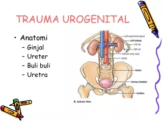
Imaging the Urogenital System. Tony Pease, DVM, MS Assistant Professor of Radiology North Carolina State University. Reading. Thrall Chapters 42-46. Prostate Gland. Not visible radiographically in normal dogs Enlarges with age, especially if intact Causes for enlargement Cysts

E N D
Imaging the Urogenital System Tony Pease, DVM, MS Assistant Professor of Radiology North Carolina State University
Reading • Thrall Chapters 42-46
Prostate Gland • Not visible radiographically in normal dogs • Enlarges with age, especially if intact • Causes for enlargement • Cysts • Infection • BPH • Cancer
SOMETIMES SEE THE PROSTATE GLAND IN THE PELVIC CANAL ON THE VD
Radiographic evidence of mineralization • Cancer • Chronic prostatitis • In castrated male • Cancer until proven otherwise
Mineralized prostate tumor with metastasis to medial iliac LN
Enlarged - Castrated Normal – Castrated 17 x 20 mm 11mm Tumor 26 x 32 mm
Paraprostatic cyst Two circular soft tissue opacities in the caudal abdomen
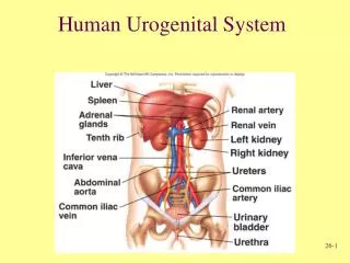
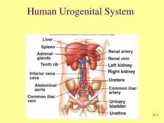
Kidneys • Retroperitoneal Organs • Right kidney cranial to the left • Left kidney more variable in location. • Size on VD Dog: 2.5 – 3.5 x length of L2 Cat: 2.4 – 3.0 x length of L2
Kidneys • Cranial pole of the right kidney • Difficult to see • Silhouettes with caudate lobe of liver • With lack of fat • Difficult to see either kidney
Mass Effect - Kidney • Kidney masses • Ventral displacement of intestine • Especially colon • A normal size kidney • Still can have disease
Bilateral renal enlargement Right kidney Right kidney Left kidney Left kidney • Lymphoma • FIP • Hydronephrosis
Small Kidneys + Mineralization Line is 2.4x L2
Small Kidneys + Mineralization Line is 2.4x L2
Excretory Urography • Improves morphologic assessment • Poor test of function • Iodinated water-soluble contrast medium • Blood flow • Glomerular filtration • Tubular reabsorption of water
Glomerulus Afferent arteriole I I I I I I I I I I I I I I I I I I I I I I I I I I I I Tubule I I I I I I I H20 I I H20 I I I I I I I I I Iodine concentration increases in tubule as H2O is resorbed
Excretory Urogram • Contrast medium • Ionic water soluble • Non-ionic if patient is compromised • Contraindications • Azotemia + dehydration • Pheochromocytoma • Multiple myeloma • Prior allergic reactions to C.M.
Excretory Urogram Standard Protocol • Survey radiographs • Inject contrast medium rapidly • 400mg Iodine per pound • Approximately 1 ml per pound • Usually do not exceed 50 ml
Projections • Immediate VD radiograph • Vascular phase • Lateral and VD radiographs at 5 minutes • Nephrogram and pyelogram phases • Lat and VD radiographs at 20 and 40 min. • Urogram phase
T=0 T=5 T=20
Urinary tract rupture Left ureter rupture Dilated left renal pelvis & proximal ureter Bladder rupture Loss of serosal detail.
Pyelonephritis • Blunt diverticulae • Dilated pelvis
Misshapen & Pyelonephritis Hydronephrosis
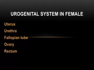
Vaginography Gartner’s duct
Technique • General anesthesia • Foley catheter placed inside labia • Labia clamped to seal outlet • Contrast medium infused • Resistance will often be encountered
Normal Retrograde Vaginogram Vagina Vestibule Urethra
Ectopic Ureters Vagina Ectopic ureters Vestibule Urethra
Urinary Bladder • Easier to evaluate with ultrasound • Very few radiographic urinary tract studies are performed • If suspect bladder problem • Carefully consider relative merits • Radiography versus ultrasonography
Urinary Bladder • To identify significant bladder problems in survey radiographs is unusual • Stones • Gas in wall; rare
Urethral Calculi “BUTT SHOT” useful to assess entire urethra
Emphysematous cystitis • Diabetes (glucosuria) with secondary bacterial infection
Urinary Bladder • Contrast Examinations • Positive contrast cystogram • Put contrast medium in bladder • Negative contrast cystogram • Rarely used • Don’t use room air; use CO2 • Double contrast cystogram • Useful for mucosa assessment
Cystography Double Contrast Cystogram Positive Contrast Cystogram
Urinary Bladder – Contrast • Double Contrast Cystogram necessary to evaluate bladder mucosa. • Assess: • Serosal margin • Mucosal margin • Bladder wall thickness • Luminal filling defects
Positive contrast cystography • Looking for rupture
Air Embolism • More common in cats • Especially those with hematuria Air in C.V.C. Air around bladder neck
Air Embolism Air in P.A. Air in R.V.
What to do! • Put animal in left lateral recumbency • Elevate the tail • This makes the right ventricle the highest point • Traps air before getting to lungs
What not to do! • Scream • Panic • Cry