Download
1 / 45
450 likes | 626 Views
Skull, Brain and Cranial Nerves. streetanatomy.com. www.uberreview.com. Skull. pg 752. Part of Axial Skeleton Cranial bones = cranium Enclose and protect brain Attachment for head + neck muscles Facial bones =framework of face Form cavities for sense organs

E N D
Skull, Brain and Cranial Nerves streetanatomy.com www.uberreview.com
Skull pg 752 • Part of Axial Skeleton • Cranial bones = cranium • Enclose and protect brain • Attachment for head + neck muscles • Facial bones =framework of face • Form cavities for sense organs • Opening for air + food passage • Hold teeth • Anchor face muscles
Cranial - 8 Frontal Occipital Sphenoid Ethmoid Parietal (2) Temporal (2) Facial – 14 Mandible Maxilla (2) Zygomatic (2) Nasal (2) Lacrimal (2) Palatine (2) Vomer Inf. Nasal Conchae(2) Cranial and Facial Bones
Bones of Skull pg 769 • Flat bones: thin, flattened, some curve • Sutures: immovable joints joining bones • Calvaria = Skullcap =Vault • Superior, Lateral, Posterior part of skull • Floor = Base • Inferior part of skull • 85 openings in skull • Spinal cord, blood vessels, nerves • Foramina, meatus, canal, fissure, notch pg 774
Cranial Fossae • Created by bony ridges • Supports, encircles brain • 3 Fossae • Anterior • Middle • Posterior pg 774
Other Cavities of Skull • Orbits • Nasal Cavity pg 854 • Middle Ear • Inner Ear pg 764
Skull through Life • Ossifies late in 2nd month of development • Frontal + Mandible start as 2 halves-then fuse • Growth of Skull • ½ adult size by age 9 months • ¾ adult size by 2 years • 100% adult size by 8-9 years • Face enlarges between ages 6-13 years
Skull bones separated by unossified membranes = Fontanels Allow compression of skull during delivery Allows rapid growth of brain Mostly replaced w/bone after 1st year Fetal Skull www.aafp.org
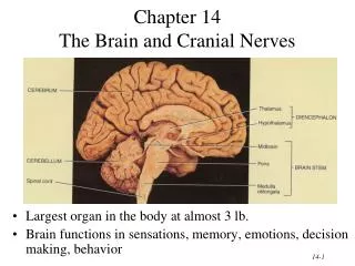
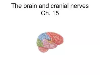
The Brain Page 788 • 4 Parts • Cerebrum • Diencephalon • Brain Stem • Pons • Medulla • Midbrain • Cerebellum • Gray matter surrounded by White matter
Meninges: 3 membranes around brain and spinal cord pg 782 • Made of Connective tissue • Functions • Cover, Protect CNS • Enclose, protect blood vessels supplying CNS • Contain CSF • 3 Layers • Dura Mater (external) • Arachnoid Mater (middle) • Pia Mater (internal)
Meninges (continued) • Dura mater • Strongest, 2 Layers, Fibrous Connective Tissue • Periosteal layer (Periosteum): External/superficial layer • Meningeal layer: Internal/deep layer • Layers fused except around dural sinuses (venous blood filled internal jugular vein) pg 769
pg 783 Extensions of Dura Mater • Partitions: limit movement of brain • Falx Cerebri–vertical, between cerebral hemispheres • Falx Cerebelli-vertical, between cerebellar hemispheres • Tentorium Cerebelli–horizontal, between cerebrum and cerebellum
Meninges pg 785 • Arachnoid Mater • Middle layer • Subarachnoid Space-between arachnoid mater and pia mater (contains most of CSF, blood vessels) • Arachnoid Villi- projections of arachnoid mater through dura into superior sagittal sinus, act as valves to help CSF pass into dural sinuses
Meninges (continued) pg 785 • Pia Mater • Innermost layer • Delicate, highly vascular • Clings directly to brain tissue, dips into convolutions
Ventricles Page 788 • Expansions of brain’s central cavity • Lined with Ependymal Cells • Filled with CSF (cerebrospinal fluid) • Ventricles continuous w/each other + central canal of spinal cord
Ventricles (continued) health.howstuffworks.com • Lateral Ventricles (#1+2) • Cerebral Hemisphere • Separated by Septum Pellucidum • Third Ventricle • Diencephalon • Interventricular Foramen: connects to lateral ventricle • Fourth Ventricle • Hindbrain • Cerebral Aqueduct: connects 3rd and 4th ventricles • Connects to central canal of spinal cord & medulla • 3 openings connect 4th to subarachnoid space • 2 lateral apertures • 1 median aperture
Cerebrospinal Fluid health.allrefer.com • Liquid cushion for brain and spinal cord • Nourishes brain • Removes waste • Conducts chemical signals between parts of CNS (e.g. hormones) • Forms as a filtrate of blood in choroid plexuses
www.daviddarling.info Choroid Plexuses • Choroid Plexuses: groups of capillaries surrounded by ependymal cells • Made of sodium, chloride ions, proteins, glucose, O2
Flow of CSF faculty.washington.edu • Formed in Choroid plexuses • Through Ventricles • Into Subarachnoid space & central canal from 4th ventricle • Through Arachnoid Villi into Superior Sagittal Sinus • Into Internal Jugular Vein
Composed of gray and white matter Different organization than in the spinal cord Cortex: external sheets of gray matter in cerebrum & cerebellum Nuclei: deep masses of gray matter surrounded by white matter Organization of the Brain
Cerebrum • “Executive Suite” of nervous system • Self-awareness, initiate + control voluntary movements, communicate, remember, understand • Made of Gray matter, White matter, Basal gangli (nuclei) • Most superior region • Covers diencephalon + top of brain stem like mushroom cap
Fissures and Grooves Fissures – deepest Transverse cerebral fissure Separates cerebral hemisphere from cerebellum Longitudinal fissure Separates R and L cerebral hemispheres Sulci Grooves on surface Gyri Ridges of brain tissue between the sulci pg 788 Cerebral Hemispheres pg 785
Cerebral Hemispheres: pg 788 • Each hemisphere divided into 5 lobes • Frontal • Parietal • Occipital • Temporal • Insula • Created by deep sulci • Functional areas: motor, sensory • Associative areas: integrate
Diencephalon pg 788 • Surrounded by cerebral hemispheres • Made of 3 Paired Structures • Thalamus • Communicates sensory info of cerebral cortex • Hypothalamus • Regulates many body activities • Emotion, sleep, memory, etc. • Pituitary Gland • Growth Hormone • Thyroid Stimulating Hormone • Epithalamus • Pineal Gland-melatonin
Brainstem: pg 788 • Medulla Oblongata, Pons, Midbrain • Passage of all signals between spinal cord and brain Midbrain Pons Medulla oblongata
Brainstem: Medulla Oblongata • Regulates several basic physiological functions • Heartbeat (rate and force) • Blood pressure (vasoconstriction/dilation of arteries) • Breathing (rate and depth) • Others: speech, coughing, sneezing, salivation, swallowing, gagging, vomiting, sweating • Attachment of CN IX, X, XI, XII
Brainstem: The Pons • Contains many tracts carrying signals: • from cerebrum to cerebellum & medulla • up to thalamus • between right and left hemispheres of cerebellum • from brainstem to cerebellum • Attachment of CN V, VI, VII, VIII
Brainstem: Midbrain • Carries signals • Between higher and lower brain centers • From cerebellum to cerebral cortex • Visual and Auditory reflex centers • Somatic motor • Attachment for CN III, IV
Cerebellum pg 788 • Smooths + coordinates body movements directed by other parts of brain • 2 Cerebellar Hemispheres • Functions • Information on equilibrium • Movement of neck, trunk, limbs • Information from Cerebral cortex
Protects brain from blood-borne toxins (e.g. urea, food toxins, bacteria) Endothelium of brain capillaries are loaded with tight junction to decrease permeability Not complete protection, some things still have to get through (e.g. fat-soluble molecules can pass through) Blood Brain Barrier
Arteries External carotid arteries and branches Tissues of head & face, skin, muscles Middle meningeal a. = brain Boxers! Internal carotid arteries and branches Opthalmic a. = Eye & Orbits Ant & Middle Cerebral arts = Cerebrum Vertebral arteries Posterior brain Vertebrae & Cervical Spinal Cord Branches form Cerebral Arterial Circle = Anastomosis Blood Supply to Brain pg 784 pg 790
Veins Dural sinuses Intracranial-receive blood from veins in brain, bring to Internal Jugular Internal jugular Drains brain External jugular Drains scalp and face (superficial) Vertebral Drains cervical vertebrae, cervical spinal cord, small neck muscles pg 823 Blood Supply to the Brain pg 828
pg 803 Cranial Nerves • 12 Pairs: I-XII • Numbered Anterior to Posterior • Attach to inferior surface of brain • Exit brain through foramina in skull • I + II attach to Forebrain (cerebrum + diencephalon) • III-XII attach to Brainstem (midbrain, pons, medulla) • Only X goes beyond the head-neck
Foramina serving Cranial Nerves • You must know what foramina each CN leaves the skull through • (refer to lab manual)
How to Remember CN I-XII Oh! Oh! Oh! To Touch And Feel Very Good Velvet! Ah Heaven!
I Olfactory (oh) II Optic (oh) III Oculomotor (oh) IV Trochlear (to) V Trigeminal (1-3) (touch) VI Abducens (and) VII Facial (feel) VIII Vestibulocochlear (very) IX Glossopharyngeal (good) X Vagus (velvet) XI Accessory (ah) XII Hypoglossal (heaven)
Motor vs. Sensory Nerves • Sensory = Afferent • Send nervous impulse from sensory receptors to brain to bring in information • e.g. pressure, temperature, pain • Motor = Efferent • Send nervous impulses from brain to body to accomplish an action • e.g. movement of a muscle, activation of a gland
Sensory Nerves • Sensory = Afferent • Visceral Sensory (sensory innervation of viscera) • stretch, pain, temp., chemical changes, irritation in viscera • Special: taste • Somatic Sensory (sensory innervation of outer part body) • touch, pain, pressure, vibration, temp. in skin, body wall, limbs • Special: hearing, equilibrium, vision, smell
Motor Nerves • Motor Nerves • Visceral Motor(motor innervation muscle in viscera + glands) • innervation smooth + cardiac muscle, glands • Branchial Motor(motor innervation of pharyngeal arch m.) • facial expression, pharyngeal constrictors, suprahyoid, sternocleidomastoid, trapezius • Somatic Motor(motor innervation of skeletal muscle) • innervation of skeletal muscles (except pharyngeal arch m.)
Mnemonic for CN Function • Some (CN I) • Say (CN II) • Marry(CN III) • Money (CN IV) • But (CN V) • My (CN VI) • Brother (CN VII) • Says (CN VIII) • Big (CN IX) • Brains(CN X) • Matter (CN XI) • Most! (CN XII) • S = Sensory functionM = Motor function • B = BOTH (Sensory and Motor function)
Cranial Nerve Function IOlfactory--------Sensory--smell II Optic-------------Sensory--vision III Oculomotor----Motor----extrinsic eye muscles IV Trochlear-------Motor----extrinsic eye muscles V Trigeminal V1Opthalmic-----Sensory-cornea, nasal mucosa, face skin V2 Maxillary------Sensory-skin of face, oral cavity, teeth V3Mandibular---Motor-muscles of mastication ---Sensory-face skin, teeth, tongue (general)
Distribution of sensory innervation to skin of face from CN V pg 819 CN V = Trigeminal V1 = Opthalmic V2 = Maxillary V3 = Mandibular
Cranial Nerves (continued) VIAbducens--------------Motor-----eye abduction muscles VII Facial-------------------Sensory---part of tongue (taste) -------------------Motor------muscles of facial expression VIII Vestibulocochlear---Sensory----hearing, equilibrium IX Glossopharyngeal----Motor------stylopharyngeus muscle ----Sensory----tongue (gen & taste), pharynx X Vagus------------------Motor-------pharynx, larynx -------------------Sensory----pharynx, larynx, abd. organs XI Accessory-------------Motor------trapezius, sternocleidomastoid XII Hypoglossal----------Motor-------tongue muscles
Summary of Functional Groups • Purely Sensory = I, II, VIII • Primarily Motor = III, IV, VI, XI, XII • Mixed = V, VII, IX, X • Parasympathetic Fibers = III, VII, IX, X (Division of Autonomic NS = Visceral Motor)
Parasympathetic Fibers pg 80 • CN III = Oculomotor • Contracts Iris (controls pupil) • Contracts Ciliary Muscle (controls lens) • CN VII = Facial • Innervates Salivary glands (mandibular & sublingual) • Innervates Lacrimal gland • CN IX = Glossopharyngeal • Innervates Parotid Salivary gland • CN X = Vagus • Innervates thoracic & abdominal viscera