Download

1 / 36
390 likes | 623 Views
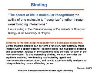
Binding Quantification with Thermophoresis. Measurements. Speaker: Christian Niederauer. Outline. Protein-Protein Interactions Grb2 Dimerization β- Lactamase TEM1 Binding to its I nhibitor BLIP Protein-Peptide Interaction: AMA1 and RON2

E N D
Binding QuantificationwithThermophoresis Measurements Speaker: Christian Niederauer
Outline • Protein-Protein Interactions • Grb2 Dimerization • β-Lactamase TEM1 Binding toitsInhibitor BLIP • Protein-Peptide Interaction: AMA1 and RON2 • Analyzing GPCR Membrane Proteins NTS1 & A2aR • Cooperative binding: Synaptotagmin
Outline • Protein-Protein Interactions • Grb2 Dimerization • β-Lactamase TEM1 Binding toits Inhibitor BLIP • Protein-Peptide Interaction: AMA1 and RON2 • Analyzing GPCR Membrane Proteins NTS1 & A2aR • Cooperativebinding: Synaptotagmin
Protein-Protein Binding: Grb2-Grb2Dimerization Grb2 dimers may control activity of FGFR2 Dimerization of Grb2 is analyzed Dimerizationaffinitytoo high for ITC approach DLS yieldsreliableresultswecancompareto MST!
Fixed ConcentrationofLabeledGrb2 KD=0.65 ± 0.08 µM monomer dimer MST allowsusageofproteinconcentrationsfarbelowactualKD DLS: • Monomer predominantupto 0.4 µM • DLS: Dimer predominantfrom 0.4µM upwards consistentwith MST measurements
Outline • Protein-Protein Interactions • Grb2 Dimerization • β-Lactamase TEM1 Binding toits Inhibitor BLIP • Protein-Peptide Interaction: AMA1 and RON2 • Analyzing GPCR Membrane Proteins NTS1 & A2aR • Cooperativebinding: Synaptotagmin
Protein-Protein Binding: β-Lactamase TEM1 -BLIP TEM-1: anti-antibioticenzyme BLIP: inhibits TEM-1 Contributionofdifferent aminoacids? Investigation withproteinmutants
Fixed ConcentrationofLabeledwt-TEM-1 wt-BLIPistitrated • KD=3.8 ± 1 nM(check: 3.2 ± 0.6nM)
Fixed ConcentrationofLabeledwt-TEM-1 W112A-BLIPistitrated • KD=0.5 ± 0.1 µM (check: 0.36 µM) position112 tryptophanalanine
Fixed ConcentrationofLabeledwt-TEM-1 W150A-BLIPistitrated • KD=1.7 ± 0.4 µM (check: 3.8 ± 0.6 µM) position150 tryptophanalanine
Mirrored Assay: Reversed Titration Protocol Fixed ConcentrationofLabeledwt-BLIP
MirroredAssay: Reversed Titration Protocol Fixed ConcentrationofLabeledwt-BLIP • wt-TEM: KD= 4.8 ± 1.7 nM (check:KD=3.8 ± 1 nM) • wt-TEM in lysate: KD=10 ± 4 nM R243A-TEM: KD= 190 ± 50 nM position243 argininealanine
Outline • Protein-Protein Interactions • Grb2 Dimerization • β-Lactamase TEM1 Binding toits Inhibitor BLIP • Protein-Peptide Interaction: AMA1 and RON2 • Analyzing GPCR Membrane Proteins NTS1 & A2aR • Cooperativebinding: Synaptotagmin
Protein-Peptid Binding: AMA1 –RON2 • Prakash Srinivasa, et al. : Binding of Plasmodiummerozoite proteins RON2 and AMA1 triggers commitment to invasion, Proceedingsofthe National Academy ofSciences 2011
Protein-Peptid Binding: AMA1 –RON2 • Plasmodium falciparum invades red blood cells and causes malaria • invasion is critically dependend on RON2-AMA1-interaction • quantification of AMA1-RON2 binding via MST • RON2 has two cysteine residues forming a disulfide bridge: • essential for binding to AMA1 • RON2 mutation/alkylation experiments show abolished binding unlabeled
Fixed Concentration of Labeled RON2 MST: Single binding event with KD= 28 ± 2 nM Check: SPR withKD = 13 ± 1 nM
Reverse Assay: Fixed Conc. ofAMA1-NT647 MST: two binding events!
Closer Look at the Two Binding Events • each half of datapoints is fitted • high affinity KD= 62 ± 16 nM • lowaffinityKD= 1.4 ± 0.2 µM • fit of equation including two events • high affinity KD= 81 ± 21 nM • low affinity KD= 1.2 ± 0.1 µM high affinity KD varies because the fit is restricted to the first half of data saturation plateau is missing because low affinity event is superimposing
Protein-Peptid Binding AMA1 –RON2 • shows importance to perform the assay in both ways • AMA1-titration only shows high-affinity binding event • RON2-titration reveals low-affinity binding event • high affinity KD has to be inferred from AMA1-titration • unlabeled RON2 peptide might have two forms in solution: with and without disulfide bond (cyclized and linear form)
Outline • Protein-Protein Interactions • Grb2 Dimerization • β-Lactamase TEM1 Binding toits Inhibitor BLIP • Protein-Peptide Interaction: AMA1 and RON2 • AnalyzingGCRP Membrane Proteins NTS1 & A2aR • Cooperativebinding: Synaptotagmin
Analyzing GCRP Membrane Proteins largest class of membrane proteins encoded in the human genome target of approximately 40% of all modern medicinal drugs! • seven transmembrane helices • connected by flexible loops • great structural diversity at the extracellular ligand binding site
GCRP: Neurotensin Receptor 1 (NTS1) Neurotensin: found in neural tissue and possibly related to mental disorders binding of Neurotensin to its receptor examined influence of NTS1 antagonist SR48692 interesting label-free approach possible due to Trp in NTS1
Binding of NTS to NTS1 Titrating NT to unlabeled NTS1: KD 20nM Titrating NTS1 to labeled NT: KD= 21 ± 20 nM high error: NTS1 amount was limited saturation could not be reached • SPR check: • 1-2 nM for both unlabeled • 7 ± 3 nMfor TAMRA-labeledNeurotensin • 1.4 nMfor Cy5-labeled Neurotensin KD dependence on fluorophore
Effect of Antagonist on NTS1 Label-free SR48692 titrated to NTS1: KD= 15 ± 11 nM(literature check: 3-10 nM) Competition Assay: SR titrated to pre-saturated (1µM NT) receptor: KD= 640 ± 50 nM(shift of one order of magnitude!) competition for same binding pocket
Effect of Antagonist on NTS1 • binding of SR yields opposite effect in thermophoretic depletion • NT promotes conformational changes in NTS1 (100 kDa) • SR locks NTS1 in an inactive conformation different hydration shell thermophoretic properties change
Adenosin A2A Receptor • A2aR is regulates myocardial blood flow • binding is observed with label-free MST: orthosteric antagonists: caffeine (19µM) theophylline (14µM) ZM241385 (1.2nM) allosteric ligand: amiloride (12µM)
A2aR Binding to Orthosteric Antagonists • Caffeine: 40± 17 µM • Theophylline : 5± 2µM • ZM241385: 43nM comparably small change in thermophoretic mobility
Influence of Amiloride to A2aR Binding Caffeine @250µM amiloride 40µM 84 ± 10µM Theophylline@250µM amiloride 5 nM 27 ± 6µM Amiloride (KD = 52 ±7µM) induces conformational change: receptor activity and hydration shell change • thermophoretic mobility increases strongly and changes signs MST can also be used to investigate allosteric binding effects!
Outline • Protein-Protein Interactions • Grb2 Dimerization • β-Lactamase TEM1 Binding toits Inhibitor BLIP • Protein-Peptide Interaction: AMA1 and RON2 • Analyzing GPCR Membrane Proteins NTS1 & A2aR • Cooperativebinding: Synaptotagmin
Synaptotagmin (syt1) Ca2+-sensor involved in neurotransmitter-release
Cooperative Binding: Synaptotagmin Synaptotagmin binds to both Ca2+ and membranes with incorporated PIP2 • Ca2+ • liposomes containg PIP2 • Synaptotagmin two possible pathways: A: Ca2+ binds to syt1, complex binds to liposome B: syt1 binds to liposome, Ca2+ binds to syt1
Cooperative Binding: Ca2+ andPIP2 KD,Ca=13± 3µMwith Ca2+ KD= 50± 10µM without binding of syt1 to liposomes with PIP2 rises in presence of Ca2+ reverse assay: Ca2+binding to syt1 with different PIP2 concentration
Cooperative Binding: Ca2+ and PIP2 PIP2: added directly syt1-PIP2 saturates if [PIP2] > 10µM syt1- Ca2+ saturates if [Ca2+] > 50µM
Cooperative Binding: Ca2+ and PIP2 Pathway A: syt1 saturated with Ca2+: KD for PIP2 decreases from 20µM to < 2µM Pathway B: syt1 saturated with PIP2: KD for Ca2+ decreases from 220µM to 3.3µM


![Quantification of [ 11 C]FLB 457 binding in the human brain with PET](https://cdn1.slideserve.com/3302395/slide1-dt.jpg)