Download
1 / 28
280 likes | 520 Views
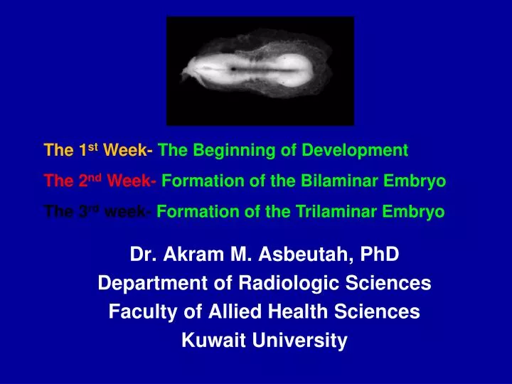
The 1 st Week- The Beginning of Development The 2 nd Week- Formation of the Bilaminar Embryo The 3 rd week- Formation of the Trilaminar Embryo . Dr. Akram M. Asbeutah , PhD Department of Radiologic Sciences Faculty of Allied Health Sciences Kuwait University. Embryology.

E N D
The 1st Week- The Beginning of Development The 2nd Week- Formation of the BilaminarEmbryo The 3rdweek- Formation of the TrilaminarEmbryo Dr. Akram M. Asbeutah, PhD Department of Radiologic Sciences Faculty of Allied Health Sciences Kuwait University
Embryology • Embryology – study of the origin and development of single individual • Prenatal period • Embryonic period – first 8 weeks • Fetal period – remaining 30 weeks
Week-1 The zygote development 2 cell zygote 4 cell zygote
Week-1 The zygote development 8 cell zygote 16 cell morula
Week-1 The zygote development Blastocyst – When zygote divides to 32 cells it Becomes known as a Blastocyst
Fertilization and the Events of the First 6 Days of Development Figure 3.3
Implantation of the Blastocyst Figure 3.4 (1 of 3)
Implantation of the Blastocyst Figure 3.4 (2 of 3)
Week 2 – The Two-Layered Embryo • Bilaminar embryonic disc – inner cell mass divided into two sheets • Epiblast and the hypoblast • Together they make up the bilaminar embryonic disc
Week 2 – The Two-Layered Embryo • Amniotic sac – formed by an extension of epiblast • Outer membrane forms the amnion • Inner membrane forms the amniotic sac cavity • Filled with amniotic fluid
Week 2 – The Two-Layered Embryo • Yolk sac – formed by an extension of hypoblast • Digestive tube forms from yolk sac • NOT a major source of nutrients for embryo • Tissues around yolk sac • Gives rise to earliest blood cells and blood vessels
Implantation of the Blastocyst Figure 3.4 (3 of 3)
Week 3 – The Three-Layered Embryo • Primitive streak – raised groove on the dorsal surface of the epiblast • Gastrulation – a process of invagination of epiblast cells • Begins at the primitive streak • Forms the three primary germ layers
Week 3 – The Three-Layered Embryo • Three Germ Layers* • Endoderm – formed from migrating cells that replace the hypoblast • Mesoderm – formed between epiblast and endoderm • Ectoderm – formed from epiblast cells that stay on dorsal surface *All layers derive from epiblast cells!
The Primitive Streak Figure 3.5e–h
The Notochord • Primitive node – a swelling at one end of primitive streak • Notochord forms from primitive node and endoderm • Notochord – defines body axis • Is the site of the future vertebral column • Appears on day 16
Formation of the Mesoderm and Notochord Figure 3.6
Neurulation • Neurulation – ectoderm starts forming brain and spinal cord • Neural plate – ectoderm in the dorsal midline thickens • Neural groove – ectoderm folds inward
Neurulation • Neurulation • Neural tube – a hollow tube pinches off into the body • Cranial part of the neural tube becomes the brain • Maternal folic acid deficiency causes neural tube defects
Neurulation • Neural crest • Cells originate from ectodermal cells • Forms sensory nerve cells • Induction • Ability of one group of cells to influence developmental direction of other cells
The Mesoderm Begins to Differentiate • Somites – our first body segments • Paraxial mesoderm • Intermediate mesoderm – begins as a continuous strip of tissue just lateral to the paraxial mesoderm
The Mesoderm Begins to Differentiate • Lateral plate – most lateral part of the mesoderm • Coelom – becomes serous body cavities • Somatic mesoderm – apposed to the ectoderm • Splanchnic mesoderm – apposed to the endoderm
Day 19 The neural tube is seen along with somites on either side of it. Somites - zipper-like motion of the neural tube closing together, three pairs of small bumps form on either side of the closure. they will form the skeleton and the major muscles of the body. Thirty-eight pairs of somites will line the neural tube within 2 weeks.