Download
1 / 37
791 likes | 2.21k Views
Schistosoma Parasites. Called Blood Flukes. Adult Flukes live in blood vessels of infected Humans. S.haematobium. S.mansoni. S.japonicum. D.H: Man R.H: None. D.H: Man R.H: Rodent, Monkeys. D.H: Man R.H: Domestic animals. Habitat . Blood vessels. Definitive Host . Man .

E N D
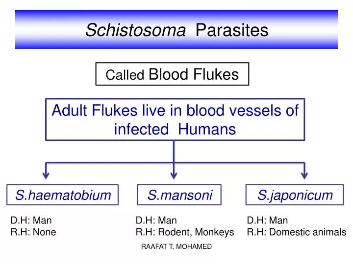
Schistosoma Parasites Called Blood Flukes Adult Flukes live in blood vessels of infected Humans S.haematobium S.mansoni S.japonicum D.H: Man R.H: None D.H: Man R.H: Rodent, Monkeys D.H: Man R.H: Domestic animals RAAFAT T. MOHAMED
Habitat Blood vessels Definitive Host Man Inferior mesenteric plexus of veins Superior & Inferior mesenteric plexus of veins Vesical & pelvic plexus of veins Where adult S.haematobium live Where adult S.mansoni live Where adult S.japonicum live RAAFAT T. MOHAMED
Schistosoma differs from other trematodes in: • Has separate sexes. • Lives in blood. • Has non-operculated eggs. • Has no Redia stage. • Has no encysted metacercaria. • Common species affecting man: • S. mansoni, S. haematobium, • S. japonicum, S. intercalatum and • S. mekongi. RAAFAT T. MOHAMED
Recent discovery: Theodore Bilharz: discovered the wormsand their relation to the disease in 1851 in Cairo. Manson: (1893): Discovered that there are 2 different species of the parasite in Egypt. Leiper (1915- 1918): Discovered the relation to snail intermediate hosts and the mode of infection. McDonagh (1918) Used tartar emetic for treatment RAAFAT T. MOHAMED
Adult Morphology Sexes:separate Female:cylindrical Male: flattened, folded (gynaecophoric canal). Suckers:oral & ventral (smaller in females). Digestive system: No muscular Pharynx Intestinal ceaca reunite in a single caecum. RAAFAT T. MOHAMED
Male RAAFAT T. MOHAMED
Female RAAFAT T. MOHAMED
Egg RAAFAT T. MOHAMED
Miracidium Snail I. H S. Haematobium Bulinustruncatus S. mansoni Biomphalariaalexandrina S. japonicum Oncomelania RAAFAT T. MOHAMED
Infective stage (Cercaria): Formed of body and forked tail (furcocercous) 5-6 pairs of penetration glands. Skin penetration is aided by: 1-Proteolytic enzymes. 2-Surface tension of drying water. 3-Movement of the tail RAAFAT T. MOHAMED
Worms mate in the liver Worms in their habitat Female lays eggs Schistosomulum reaches circulation Miracidium infects snail i.h. Eggs in urine or stools Infection in contaminated water Cercarial shedding Miracidia hatch RAAFAT T. MOHAMED
Life cycle RAAFAT T. MOHAMED
Development of Schistosomainside the body of infected human Aorta II Systemic circulation Venous circulation Liver Schistosomula ♀ Vesicalplexus ♂ S.haematobium I Portal vein Inf. mesenteric plexus Cercariae penetrates human skin III S.mansoni IV Direction of venous blood flow In urine or stool RAAFAT T. MOHAMED
Pathogenesis and Clinical Picture There are 4 progressive stages: I- Stage of invasion (1-4 days) Local dermatitis, irritation. Papular rash. RAAFAT T. MOHAMED
(3-4 weeks) II- Stage of migration By schistosomula Metabolic products: toxic and allergic manifestations as urticaria, fever, headache, muscle pain. In the lung: verminous pneumonitis, minute haemorrhage cough, haemoptysis. In the liver: enlarged and tender. RAAFAT T. MOHAMED
III- Stage of egg deposition and extrusion (acute stage: 1-2 month) A- Eggs are deposited in the venous plexus The patient may complain of: Generalized malaise, fever, rigors, urticaria, abdominal pain and liver tenderness. Urticaria RAAFAT T. MOHAMED
Occurs mainly in S.japonicum infection Katayama Syndrome Due to: 1- ♀lays large number of eggs 2- greater proximity to the liver Soluble egg antigens are released in blood stream immune complex Circulate Blood vessel antibodies Deposited in the tissues Tissue damage The patient suffers from: Fever, chills, diarrhoea, generalized lymphadenopathy Thus also called acute toxoemic schistosomiasis RAAFAT T. MOHAMED
1-Pressure within the venule B- Stage of egg extrusion: 2-Effect of the spine Eggs escape from the veins to the perivascular tissue due to: 3-oozing lytic secretion of the miracidium within the egg Damage & haemorrhage S.haematobium S.mansoni& S.japonicum In vesical& pelvic plexus of veins In mesenteric plexus of veins Egg extruded Egg extruded Bladder wall Intestinal wall Terminal haematuria, frequency of micturition, burning pain Dysentery with blood and mucus in stool RAAFAT T. MOHAMED
IV- Stage of tissue reaction (chronic stage) (months – years) Shell & miracidial antigens Occurs around Schistosoma eggs deposited in various tissues. Granuloma formation in tissue Granuloma develops Attract inflammatory cells Deposition of fibrous tissue Damage of affected organ and its fibrosis miracidium Inflammatory cells Egg shell Delayed-type hypersensitivity Loss of its function RAAFAT T. MOHAMED
In normal condition In S.haematobium infection hydronephrosis Normal mucosa of colon hydroureter Fibrous tissue Egg trapped in bladder wall Polyps in the colon in S.mansoniinfection Stone formation Cancer bladder may occur RAAFAT T. MOHAMED
IV- Stage of tissue reaction (chronic stage) In S.haematobiuminfection In S.mansoniinfection Eggs fail to fix to venule wall are swept by blood Eggs fail to fix to venule wall are swept by blood 3 3 2 Eggs trapped in Intestinal wall Eggs trapped in Bladder wall 2 1 Eggs extruded in stool Eggs extruded in urine 1 Cause Embolic lesions Cause Embolic lesions RAAFAT T. MOHAMED
Embolic Lesions in the Liver Hepatosplenomegaly Portal hypertension Oesophagealvarices Ascites. RAAFAT T. MOHAMED
Embolic Lesions in the lung Bilharzialcor-pulmonale Pulmonary hypertension In Schistosoma infection In Normal condition Trapped eggs Dilated right ventricle Thickened wall Enlarged right ventricle Cor-pulmonale = right-sided heart failure Means: enlargement of right ventricle due to high blood pressure in the arteries of the lung Embolic lesions may occur in any other organ. RAAFAT T. MOHAMED
Diagnosis of Schistosomiasis Clinically Laboratory Radiological imaging Endoscopy (suggestive) 1- History of contact with infected water. 1- Detection of eggs in urine or stool 2- Clinical picture according to stage of infection. 2- Blood examination 3- Serological tests RAAFAT T. MOHAMED
I- Laboratory Diagnosis 1- Direct parasitological methods: - Detection of S.haematobiumeggs in urine. - Detection of S.mansoni eggs in stool by direct smear method or by concentration method. - Test for viability. - Kato thick faecal smear: for egg counting to assess the intensity of infection - Rectal swab S: 140X60 µ 150X60 µ S: Terminal spine Oval, thin shell Lateral spine C: Translucent C: Miracidium RAAFAT T. MOHAMED
I- Laboratory Diagnosis 2- Blood examination: 1- Egg extrusion Iron deficiency anaemia Anaemiadue to 2- Hypersplenism Haemolytic anaemia Eosinophilia, leucocytosis. 3- Serological Tests: (Immunodiagnostic Tests) Detection of anti-Schistosoma antibodies or antigen in patient’s serum a- IHAT (Indirect Haemagglutination test) Sensitized sheep RBCs Latex particles +ve -ve RAAFAT T. MOHAMED
b- ELISA (Enzyme-linked immunosorbent assay) Coating with Schisto Ag Patient’s serum Ab linked to enzyme substrate Ab detected wash wash wash Coating with anti-Schisto MAb Patient’s serum Ab linked to enzyme substrate Ag detected wash wash wash c- IFAT (Indirect Fluorescent Antibody test) Ab detected Patient serum Schisto Ag wash wash RAAFAT T. MOHAMED
II- Radiological imaging S.haematobium infection S.mansoni infection Calcified bladder with hydroureter Stenosed ureters and hydronephrosis Intestinal affection RAAFAT T. MOHAMED
المناظير III- Endoscopy Cystoscopy in S.haematobium Colonoscopy, sigmoidoscopy in S.mansoni Done in chronic cases to detect lesions and take biopsies RAAFAT T. MOHAMED
Treatment Praziquantel (Biltricide): For all schistosome species. Dose:40 mg/kg single oral dose Oxamniquine (vancil): For S. mansoni Dose: 30 mg/kg for 2 days Metriphonate: For S. haematobium only. Dose: 10mg/kg every 2 weeks for 3 doses. RAAFAT T. MOHAMED
Prevention and Control -Mass treatment. - Health Education. - Snail control: Physical methods Biological methods Chemical methods X Balanites aegyptica Copper sulphate RAAFAT T. MOHAMED
Cercarial Dermatitis (Bather’s itch) A condition that occurs due to penetration of cercariae of non-human species of schistosomes the skin of man. Cercariae cannot go beyond the germinal layer. Clinical picture: Dermatitis, itching, oedema and secondary bacterial infection. Treatment: Antipruritics, antihistamincs, antibiotics. RAAFAT T. MOHAMED
Multiple Choice Questions The major cause of morbidity in schistosomiasis is: a- Migration of adult worms against blood stream. b- S.japonicum b- Maturation of worms to adult stage in the liver. c- Occurrence of Embolic lesions. d- Deposition of eggs in tissues. Kato technique is used to diagnose: a- Paragonimiasis b- Schistosomiasis mansoni c- Schistosomiasis haematobium d- Fascioliasis Bather’s itch occurs frequently with: b- S.mansoni a- S.haematobium d- non human schistosomes RAAFAT T. MOHAMED
Multiple Choice Questions Katayama syndrome: a- occurs most frequently in S.haematobiuminfection. b- occurs most frequently in S.mansoniinfection. c- occurs most frequently in S.japonicuminfection. d- occurs in chronic schistosomiasis. RAAFAT T. MOHAMED
Give reasons for Occurrence of terminal haematuria in S.haematobium infection Terminal haematuria is due to active deposition of eggs by adult female S. haematobium in vesical venous plexuses. Eggs escape to the perivascular tissue and become extruded to the lumen of the urinary bladder. Powerful contraction of the bladder at the end of micturition (to squeeze the last drops of urine) leads to haemorrhage (terminal haematuria). Egg passes lumen of urinary bladder RAAFAT T. MOHAMED
Give reasons for Occurrence of Bilharzialcor-pulmonale in schistosomiasis Many eggs that fail to be fixed to walls of venules are swept by blood to reach various organs as the lungs where they form granulomas and fibrosis with obliteration of flow resulting in pulmonary hypertension and right-sided heart failure. Swept by blood Heart Lung RAAFAT T. MOHAMED