Download
1 / 1
10 likes | 108 Views
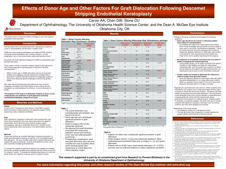
Carver AA, Chen GW, Stone DU Department of Ophthalmology, The University of Oklahoma Health Science Center, and the Dean A. McGee Eye Institute Oklahoma City, OK. Effects of Donor Age and Other Factors For Graft Dislocation Following Descemet Stripping Endothelial Keratoplasty. Conclusions.

E N D
Carver AA, Chen GW, Stone DU Department of Ophthalmology, The University of Oklahoma Health Science Center, and the Dean A. McGee Eye Institute Oklahoma City, OK Effects of Donor Age and Other Factors For Graft Dislocation Following Descemet Stripping Endothelial Keratoplasty Conclusions Disclaimer Results Findings in this group of patients would support the following guidelines: • Donor age should not be a factor in selecting suitable donor tissue for transplantation. • Our findings also support the recent report of the Cornea Donor Study Investigator Group that did not find an effect of donor age on outcomes of penetrating keratoplasty.12 While the recent report by Terry and colleagues did not report a statistically significant difference in dislocation rates related to storage time,5 their reported times were shorter than those in this cohort, possibly explaining the discrepancy. • Be cognizant of iris anatomy and intraocular lens status in patient counseling and surgical planning. • During DSEK surgery, an air bubble is placed in the anterior chamber to help stabilize and retain the newly placed endothelial graft, and if the patient has a superior iris defect that will allow the air bubble to be displaced posteriorly, we hypothesize that the graft is much more likely to dislocate. • Further studies are needed to determine the influence of death-to-surgery time and other factors. • Given the apparent difference in dislocation rates with grafts that were transplanted 4, 5, or 6 days after donor’s time of death, an argument can be made for the use of tissue that has been harvested within 4 or 5 days of surgery. Regarding the graft dislocation rate of 26.3%, these surgeries were attended by one surgeon, but in a high percentage of these cases a resident or fellow was the primary surgeon. The above rate likely reflects the learning curve previously described.6 This may also be in part attributed to the more inclusive definition of graft dislocation, including grafts with a fluid cleft that resolved with supine positioning in the presence of a retained anterior chamber bubble. To eliminate uncontrolled variables, this series also did not include surgeries with significant changes in technique, such as venting incisions, that have been adopted more recently. Weaknesses of this study include: • The limitations inherent to a retrospective study. • The high dislocation rate compared to a single experienced surgeon always performing the primary procedure. Future prospective studies with endpoints defined a priori will help answer these questions more definitively. The authors have no financial conflicts of interest in any of the material presented in this document. Table 1:Donor Factors Affecting Dislocation Rate (Continuous variables) Table 2: Donor Factors Affecting Dislocation Rate (Dichotomous pairings) Introduction • Corneal edema from endothelial dysfunction represents an important cause of visual disability and decrease in quality of life.1 • DSEK has many potential advantages over traditional PKP including shortened visual rehabilitation time, minimal change in corneal astigmatism, and a more anatomically stable post-operative result.2 • At present, the most significant drawback to DSEK is postoperative graft lenticule dislocation. • There exists a variety of potential variables related to graft adherence that is being explored in a growing body of experience reported retrospectively.2-11 • Effect of donor age on DSEK dislocation rate not yet evaluated. • O’Brien and colleagues recently reported an increased risk of dislocation in patients with abnormal lens-iris diaphragm anatomy.3 • Some have observed special challenges in performing aphakic DSEK.4 • Still, other factors that determine graft adherence have not been completely elucidated, and further studies of such criteria will only strengthen our understanding of its influence on donor dislocation in DSEK. • The purpose of this study is to determine if patient or donor cornea characteristics are predictive of graft dislocation following endothelial keratoplasty using pre-cut tissue. Materials and Methods DSEK = Descemet's Stripping Endothelial Keratoplasty Design A case-control, retrospective chart review of 194 DSEK surgeries performed using a single method by one attending surgeon (DUS) supervising various residents and fellows. Data Data collected for evaluation of dislocation rate included donor age, donor tissue endothelial cell count, donor tissue time from death to preservation, donor tissue time from death to surgery, patient demographics, etiology of endothelial dysfunction, and past ocular history (specifically extent and location of iris abnormalities and lens status). Methods Cases were defined as complete dislocations requiring reoperation or partial graft dislocations requiring re-injection of air to reestablish graft-host apposition. We also included cases where a partial dislocation was treated with supine positioning. Controls were defined as those cases with uneventful complete graft attachment. To evaluate the statistical significance between the categorical variables, two-by-two contingency tables were employed with the Fishers exact test to compute P-values. To evaluate the statistical significance of numerical mean values, the unpaired t-test was used to compute P-values. • Table 1: • The overall dislocation rate, including partial and complete, was found to be 26.3%. • Donor age was not a statistically significant predictor of graft dislocation. • Death to surgery interval was statistically significant. • We observed one graft dislocation associated with severe post-operative nausea and vomiting, which may have affected patient positioning. • Postoperative complications other than graft dislocation were rare and included one case of pupillary block which resolved quickly after releasing air to a level above the inferior pupillary margin. References 1. Boisjoly H, Gresset J, Fontaine N, Charest M, Brunette I, LeFrancois M, Deschenes J, Bazin R, Laughrea PA, Dube I. The VF-14 index of functional visual impairment in candidates for a corneal graft. Am J Ophthalmol 1999;128:38-44. 2. Terry MA. Endothelial keratoplasty: history, current state, and future directions. Cornea. 2006 Sept; 25(8):873-878. 3. O’Brien PD, Lake DB, Saw VP, Rostron CK, Dart JK, Allan BD. Endothelial keratoplasty: case selection in the learning curve. Cornea 2008;27:1114-1118. 4. Suh LH, Kymionis GD, Culberston WW, O’Brien TP, Yoo SH. Descemet stripping with endothelial keratoplasty in aphakic eyes. Arch Ophthalmol. 2008 Feb; 126(2):268-270. 5. Terry MA, Shamie N, Chen ES, Phillips PM, Hoar KL, Friend DJ. Precut tissue for Descemet’s stripping automated endothelial keratoplasty: Vision, astigmatism, and endothelial survival. Ophthalmol 2009 Feb; 116(2):248-56. 6. Price FW Jr., Price MO. Descemet’s stripping with endothelial keratoplasty in 200 eyes: early challenges and techniques to enhance donor adherence. J Cataract Refract Surg. 2006 Mar; 32:411-418. 7. Price MO, Price FW. Endothelial cell loss after Descemet stripping with endothelial keratoplasty influencing factors and 2-year trend. Ophthalmol 2008;115:857-865. 8. Koenig SB, Covert DJ. Early results of small incision Descemet’s stripping automated endothelial keratoplasty. Ophthalmol 2007;114:221-226. 9. Terry MA, Shamie N, Chen ES, Hoar KL, Phillips PM, Friend DJ. Endothelial keratoplasty: the influence of preoperative donor endothelial cell densities on dislocation, primary graft failure, and 1-year cell counts. Cornea. 2008 Dec; 27(10):1131-7. 10. Price MO, Price FW. Descemet’s stripping with endothelial keratoplasty: comparative outcomes with microkeratome-dissected and manually dissected donor tissue. Ophthalmol 2006;113:1936-1942. 11. Lee JK, Eghrari AO, Desai NR, Stark WJ, Gottsch JD. Presoaking donor corneas reduces graft detachment rates in descemet stripping endothelial keratoplasty. Am J Ophthalmol. 2009 Mar;147(3):439-441. 12. Yoo SH, Kymionis GD, Deobhakta AA, Ide Takeshi, Manns F, Culbertson WW, O’Brien TP, Alfonso EC. One-year results and anterior segment optical coherence tomography findings of Descemet stripping automated endothelial keratoplasty combined with phacoemulsification. Arch Ophthalmol 2008;126(8):1052-1055. DSEK = Descemet's Stripping Endothelial Keratoplasty, ACIOL = Anterior Chamber Intraocular Lens, PBK = Pseudophakic Bullous Keratopathy. • Table 2: • Superior iris defect was a statistically significant predictor of graft dislocation. • Death-Surgery interval > 6 days was statistically significant. When evaluating tissue harvested 6 days prior to surgery (n = 5), all 5 grafts dislocated. • Patients with an ACIOL had a trend towards dislocation (P = 0.0725), but there was not sufficient evidence to make a significant correlation. This research supported in part by an unrestricted grant from Research to Prevent Blindness to the University of Oklahoma Department of Ophthalmology For more information regarding this poster and other research studies at The Dean McGee Eye Institute visit www.dmei.org