Download
1 / 27
280 likes | 449 Views
MCB 7200: Molecular Biology. Characterization of DNA clones including: Restriction Enzyme (RE) mapping Subcloning Southerns Northerns* Westerns Hybrid-select translation* DNA sequencing* PCR. Characterization: RE mapping. Predict what would happen with a double digest.

E N D
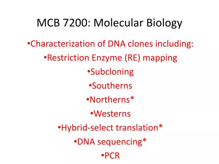
MCB 7200: Molecular Biology Characterization of DNA clones including: Restriction Enzyme (RE) mapping Subcloning Southerns Northerns* Westerns Hybrid-select translation* DNA sequencing* PCR
Characterization: RE mapping Predict what would happen with a double digest.
Characterization: Subcloning • Refers to the process of cloning smaller pieces of a large DNA cloning into another cloning vector • E.g., subcloning the individual EcoRI fragments of a partial EcoRI lambda genomic clone into plasmid vectors • Facilitates amplication and analysis of the subcloned DNA, including probe preparation
Experimental Figure 5.26 Southern blot technique can detect a specific DNA fragment in a complex mixture of restriction fragments.
Characterization: Southern blot hybridization -transfer of DNA from a gel to a membrane (e.g., nitrocellulose, nylon) -developed by Edwin Southern
Characterization: Northern blot hybridization X RNA X x salt X RNA -transfer of RNA from a gel to a membrane (e.g., nitrocellulose, nylon) -reveals mRNA size (and approximate protein size), tissue- and organ- specific expression, and kinetic patterns of expression
Experimental Figure 5.27 Northern blot analysis reveals increased expression of b-globin mRNA in differentiated erythroleukemia cells.
Experimental Figure 5.28 In situ hybridization can detect activity of specific genes in whole and sectioned embryos.
Characterization: Western blotting X Protein Enzyme reaction or X React with Antibody X x Buffer; requires electric current X -transfer of protein from a gel to a membrane (e.g., nitrocellulose, nylon) -requires the use of an electric current to facilitate transfer
Animations for Western Blotting • SDS gel electrophoresis- MCB Chapter 3 • http://bcs.whfreeman.com/lodish7e/#800911__812035__ • Western blotting or Immunoblotting- MCB Chapter 3 • http://bcs.whfreeman.com/lodish7e/#800911__812034__
Experimental Figure 3.36 SDS-polyacrylamide gel electrophoresis (SDS-PAGE) separates proteins primarily on the basis of their masses.
Experimental Figure 3.39 Western blotting (immunoblotting) combines several techniques to resolve and detect a specific protein.
Characterization: DNA Sequencing The dideoxynucleotide structure (note 3’H) is the key to dideoxy DNA sequencing
Dideoxy DNA Sequencing Animation- MCB Chapter 5 • http://bcs.whfreeman.com/lodish7e/#800911__812048__
Automated DNA sequencing Capillary electrophoresis
Pyrosequencing http://www.biotagebio.com/DynPage.aspx?id=7454
454 Sequencing (massively parallel pyrosequencing) How it Works System Workflow: One Fragment = One Bead = One Read The complete sequencing workflow of the Genome Sequencer FLX System comprises four main steps, leading from purified DNA to analyzed results. These basic steps include: 1) Generation of a single-stranded template DNA library, 2)Emulsion-based clonal amplification of the library, 3) Data generation via sequencing-by-synthesis, and 4) Data analysis using different bioinformatics tools Sample Input and Fragmentation The Genome Sequencer FLX System supports the sequencing of samples from a wide variety of starting materials including genomic DNA, PCR products, BACs, and cDNA. Samples such as genomic DNA and BACs are fractionated into small, 300- to 800-basepair fragments. For smaller samples, such as small non-coding RNA or PCR amplicons, fragmentation is not required. Instead, short PCR products amplified using Genome Sequencer fusion primers can be used for immobilization onto DNA capture beads as shown below under "One Fragment = One Bead". Library Preparation Using a series of standard molecular biology techniques, short adaptors (A and B) - specific for both the 3' and 5' ends - are added to each fragment. The adaptors are used for purification, amplification, and sequencing steps. Single-stranded fragments with A and B adaptors compose the sample library used for subsequent workflow steps. One Fragment = One Bead The single-stranded DNA library is immobilized onto specifically designed DNA Capture Beads. Each bead carries a unique single-stranded DNA library fragment. The bead-bound library is emulsified with amplification reagents in a water-in-oil mixture resulting in microreactors containing just one bead with one unique sample-library fragment. emPCR (Emulsion PCR) Amplification Each unique sample library fragment is amplified within its own microreactor, excluding competing or contaminating sequences. Amplification of the entire fragment collection is done in parallel; for each fragment, this results in a copy number of several million per bead. Subsequently, the emulsion PCR is broken while the amplified fragments remain bound to their specific beads. One Bead = One Read The clonally amplified fragments are enriched and loaded onto a PicoTiterPlate device for sequencing. The diameter of the PicoTiterPlate wells allows for only one bead per well. After addition of sequencing enzymes, the fluidics subsystem of the Genome Sequencer FLX Instrument flows individual nucleotides in a fixed order across the hundreds of thousands of wells containing one bead each. Addition of one (or more) nucleotide(s) complementary to the template strand results in a chemiluminescent signal recorded by the CCD camera of the Genome Sequencer FLX Instrument. For a detailed explanation of this reaction see Sequencing Chemistry. Data Analysis The combination of signal intensity and positional information generated across the PicoTiterPlate device allows the software to determine the sequence of more than 1,000,000 individual reads per 10-hour instrument run simultaneously. For sequencing-data analysis, three different bioinformatics tools are available supporting the following applications: de novo assembly up to 400 megabases; resequencing genomes of any size; and amplicon variant detection by comparison with a known reference sequence. See http://www.454.com/products-solutions/how-it-works/index.asp
Experimental Figure 5.23 Generation of clusters of identical DNA molecules attached to a solid support.
Experimental Figure 5.24 Using fluorescent-tagged deoxyribonucleotide triphosphates for sequence determination.
PCR Animation- MCB Chapter 5 • http://bcs.whfreeman.com/lodish7e/#800911__812049__
The Polymerase Chain Reaction (PCR) • PCR is a cloning method without a host • Thermus aquaticus, a hot spring bacterium, produces Taq polymerase • Taq polymerase, unlike E. coli DNA polymerase, is not denatured at 95°C
Figure 5.20 The polymerase chain reaction (PCR) is widely used to amplify DNA regions of known sequences.
Experimental Figure 5.21 A specific target region in total genomic DNA can be amplified by PCR for use in cloning.
Chemical synthesis of DNA(oligonucleotide synthesis using phosphoramidite chemistry)