Download
1 / 126
1.38k likes | 2.17k Views
Osteosarcoma and its Variants. Amit M. Patel, MS William Simmons, PhD. Acknowledgements. Pictures in this Power-Point was graciously provided by Dr. S. R. Patel, a practicing oncologist working at Rush Presbyterian Hospital in Chicago, IL

E N D
Osteosarcoma and its Variants Amit M. Patel, MS William Simmons, PhD
Acknowledgements Pictures in this Power-Point was graciously provided by Dr. S. R. Patel, a practicing oncologist working at Rush Presbyterian Hospital in Chicago, IL All information in these slides came from previous lecture notes on the subject at Texas A&M University and material from the Oxford Textbook of Pathology
Osteosarcoma • Definitions: • A mesenchymal malignancy (malignant spindle cells) that differentiates to produce osteoid/immature bone • Considered an osteosarcoma no matter how much osteoid is produced • Second most common primary malignant tumor of bone (first most common=multiple myeloma) • 15% of all biopsied primary bone tumors
Osteosarcoma • Definitions: • Primary Osteosarcoma: arises from the bone in the absence of a benign precursor lesion or treatment • Secondary Osteosarcoma: arises from a precursor lesion to one that is metastatic from a primary osteosarcoma • Synchronous Osteosarcoma: Lesions that affect multiple bones discovered within 6 mos of each other • Metachronous Osteosarcoma: Lesions involving multiple bones discovered more than 6 mos apart
Osteosarcoma • Definitions: • Intramedullary Osteosarcoma: Lesion arising within the medullary space of the bone (most common type) • Juxtacortical Osteosarcoma: Lesion arising on the surface of the bone in apposition to the cortex • Intracortical Osteosarcoma: Lesion arising from the cortex of the bone
Intramedullary (75%) Conventional Osteoblastic (82%) Mixed and Sclerosing Chondroblastic (5%) Fibroblastic (3-4%) MFH-like (3-4%) Osteoblastoma-like (.5%) Giant Cell-rich (.5%) Small-cell (1%) Epithelioid (.5%) Telangiectatic (3%) Well-differentiated (low grade intraosseous; 4%-5%) Juxtacortical/Surface(7-10%) Parosteal Periosteal High-grade surface Intracortical(.2%) Secondary (older population) Pagets (67-90%); Post RT (6-22%); Bone infarct; Fibrous dysplasia; Metallic implant; Osteomyelitis OS with specific syndromes Familial; Retinoblastoma; Rothmund-Thomson Syndrome; Multifocal; OI OsteosarcomaClassification
General Radiology: Plain Radiographic Presentation • Osteoid/Ossification production on X-Ray • Mixed Sclerotic and Lytic Lesion—Most common radiographic presentation • Purely Lytic • Purely Blastic
Osteosarcoma • General Pathology: • Osteoidand/or immature bone production by tumor cells • Malignant stromal cells • Graded on degree of anaplasia I-IV
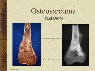
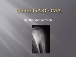
Osteosarcoma • Primary, High Grade, Intramedullary (Conventional) • About 75% of all osteosarcomas • Ages: 15-25 years (rare <6y or >60y) • Sex: Male>Female 1.5-2:1 • Sites: • Long Bones: 70%-80% • Distal Femur (40%; about twice as common as proximal tibia) • Proximal Tibia (20%) • Proximal Humerus (10-15%) • Axial Skeleton • Pelvis • Jaw
Osteosarcoma • Sites: • Metaphysis: 90% • Diaphysis: 8-10%
Telangiectatic Osteosarcoma • Tumor largely composed of cystic cavities containing necrosis and hemorrhage • ABC- like which can lead to a misdiagnosis on X-rays • Sites: Similar to conventional • Distal femur, proximal tibia, proximal humerus • Metaphyseal (90%), diaphyseal (10%)
Telangiectatic Osteosarcoma • Radiology: • Osteolytic and expansile on X-ray • Small areas of osteoid (more easily detected with CT) • Pathologic fracture (25%-30%) • MRI/CT: Fluid-fluid levels; soft tissue mass • Bone scan: Donut sign
Juxtacortical Osteosarcoma • Parosteal Osteosarcoma (65%) • Periosteal Osteosarcoma (25%) • High Grade Surface (10%)
Parosteal Osteosarcoma • Origin: Arises from outer layer of periosteum • Usually a low grade tumor with fibroblastic stroma and osteoid/woven bone • Age: 20-30 yrs; usually about a decade older than conventional osteosarcoma • Location: • Posterior distal femur metaphysis (65%) • Proximal humerus (15%); Tibia (10%); Fibula (3%) • Clinical: painless mass in posterior distal thigh; may be present for several yrs; decreased ROM of adjacent joint • Sex: Female>Male 2:1
Parosteal Osteosarcoma • Radiology: • XR: • Lobulated and ossified exophytic mass (cauliflower-like) adjacent to the cortex with a lucent cleavage plane between lesion and the cortex • Radiodense centrally • Cortical thickening • Large tumors encircle the bone • Growth may obliterate cleavage plane between lesion and cortex and will appear to have broad attachment • Invasion of the medullary canal with long standing disease
Periosteal Osteosarcoma • Low to intermediate grade bone forming sarcoma with predominant chondroblastic differentiation tumor (>90% of tumor); <2% of osteosarcomas • Origin: Arises from the inner layer of the periosteum • Age: 10-20 yrs; similar to conventional osteosarcoma • Sex: Slight male predominance • Location: Diaphysis of femur and tibia (>85%); ulna and humerus (10%)
Periosteal Osteosarcoma • Radiology: • XR: • Diaphyseal lesion on surface of bone; medullary canal is uninvolved • Saucerized cortex with chondroblastic soft tissue mass • Cortical thickening at margins of erosion (40%) • May have Codman’s triangle • Spiculated or sunburst periosteal reaction (elevates the periosteum) • Partial matrix mineralization may be seen consistent with chondroblastic nature • Rarely, intramedullary invasion
High Grade Surface Osteosarcoma • High grade osteosarcoma that develops on the surface of the bone without any medullary involvement; very rare (<1% of osteosarcomas) • Histology is the same as a conventional osteosarcoma with the same potential for mets • Age: 2nd decade • Sites: Femur (45%); Humerus (26%); Fibula (10%); arises usually on the metaphyseal surface
High Grade Surface Osteosarcoma • Radiology: • Appearance similar to periosteal osteosarcoma but matrix mineralization is similar to conventional osteosarcoma with cloudlike opacities • Broad based lesion arising on surface • Codman’s triangle; periosteal new bone • Cortical erosion/destruction but medullary cavity usually uninvolved
Low Grade Intramedullary Osteosarcoma • Intramedullary low grade fibroblastic osteoid producing sarcoma characterized by benign cytologic features of spindle cells and maturity of tumor bone • 1% of all osteosarcomas • Age: peak— 3rd decade; individual cases in 2nd decade and 50s • Sites: Metaphysis of femur and tibia most common
Low Grade Intramedullary • Radiology: • XR: • Meta-epiphyseal • Central ossification/sclerosis with expansile remodeling • Ground glass density and internal trabeculation (simulates fibrous dysplasia) • Usually no soft tissue mass and not as aggressive appearing • Usually no periosteal reaction
Intracortical Osteosarcoma • High grade osteosarcoma confined to the cortex of a long bone • Very rare; handful of cases • Age: 10-30 yrs • Sites: Diaphysis of femur or tibia • Radiology: • Intracortical lucency with surrounding sclerosis of bone • No intramedullary or soft tissue involvement • Minimal or no periosteal reaction
Conventional Osteosarcoma of Distal Femur X-Ray Codman’s Triangle Permeative Lesion Ossification in Soft Tissue Component
Conventional Osteosarcoma of Proximal Tibia Permeative Lesion with Fluffy White Ossification (sclerosis) Cortical Destruction
OsteosarcomaConventional • Radiographic Differential Diagnosis: • Ewing sarcoma • Fibrosarcoma/MFH • Chondrosarcoma • Osteomyelitis • Osteoblastoma • Giant Cell Tumor
Examples of Conventional Osteosarcomas including Gross and Microscopic Pathology
Chondroblastic Subtype of a Conventional Osteosarcoma of Distal Tibia