Download

1 / 42
470 likes | 1.04k Views
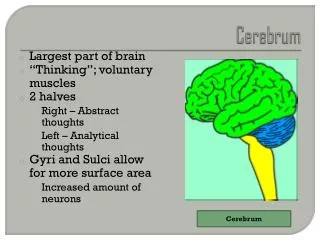
Dr. Zeenat Zaidi. CEREBRUM. Cerebrum. Corpus callosum. Largest part of the forebrain Derived from telencephalon Divided into two halves, the cerebral hemipheres The two hemispheres are separated by a deep median lingitudinal fissure which lodges the falx cerebri

E N D
Dr. ZeenatZaidi CEREBRUM
Cerebrum Corpus callosum • Largest part of the forebrain • Derived from telencephalon • Divided into two halves, the cerebral hemipheres • The two hemispheres are separated by a deep median lingitudinal fissure which lodges the falxcerebri • In the depth of the fissure, the hemispheres are connected by a bundle of fibers called the corpus callosum Right hemisphere Left hemisphere Median longitudinal fissure
Surfaces Superolateral Medial Inferior
The structure of cerebral hemipheres includes: Superficial layer of grey matter, the cerebral cortex Deeper to the cortex, axons running to and from the cells of the cortex form an extensive mass of white matter Burried within the white matter lie a number of nuclear masses (caudate, putamen, globuspallidus) collectively known as the basal ganglia Cortex Basal ganglia WM WM
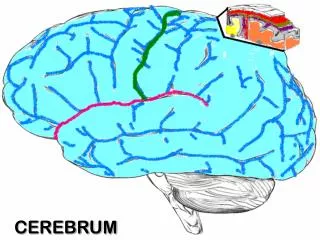
The superficial layer of grey matter is highly convoluted to form a complex pattern of ridges (gyri)and grooves (sulci) This arrangement maximize the surface area of the cerebral cortex (about 70% is hidden within the depths of sulci) S G G G
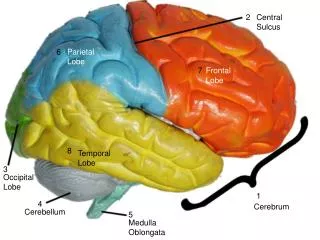
Three sulci, consistent in their position are used to divide each hemisphere into lobes. These include: Central sulcus& Lateral sulcuson the superolateral surface Parieto-occipital sulcus, on the medial surface
Each hemisphere is divide into FOUR lobes (named after overlying bones) motor function, motivation, aggression, smell and mood reception and evaluation of sensory information visual processing smell, hearing, memory and abstract thought
Functionally each hemisphere contains a ‘limbic lobe’ on the medial surface. • It is responsible for: • establishing emotional states • linking conscious intellectual functions with the unconscious autonomic functions • facilitating memory storage and retrieval
Central sulcus An uninterrupted vertical sulcus running from the lateral fissure to the median longitudinal fissure Extending for a short distance on the medial surface of the hemisphere Separates the frontal lobe from parietal lobe Lateral sulcus Deepest cleft on the lateral surface Separates the temporal lobe from the frontal and parietal lobes Parieto-occipital sulcus Separates the occipital lobe from the parietal lobe on the mdial surface F P T F P O
Frontal lobe: Precentralgyrus Superior & inferior frontal sulci divide the lobe into superior, middle & inferior frontal gyri Parietal lobe: Postcentralgyrus Intraparietalsulcus dividing the lobe into superior & inferior parietal lobules Superior , middle & inferior frontal gyri Precentral gyrus Postcentral gyrus sfs ifs Inferior parietal lobule Superior parietal lobule Intraparietal sulcus
Temporal lobe: Superior & inferior temporal sulci giving rise to superior, middle & inferior temporal gyri Insula: the gyri in the depth of lateral fissure, covered by parts of frontal, parietal & temporal lobes called the opercula (removed in lower pic.) Superior, middle & inferior temporal gyri sts its insula
Medial Surface • Sulci: Parietooccipital, Calcarine, Cingulate • Gyri: Cingulate, Parahippocampal
Histological Structure of Cerebral Cortex • Contains nerve cell bodies, dendritic arborizations, synapses, neuroglia, blood vessels • Archicortex and Paleocortex (hippocampus and parts of temporal lobe associated with olfactory functions) have three layered structure • Neocortex, generally consists of six layers, although the detailed cytological structure varies from region to region
Histological Structure of Cerebral Cortex • Layer I: few nerve cells, many processes and synaptic interactions • Layer II: many small neuron, which establish intercortical connections • Layer III: medium sized neurons giving rise to association & commissural fibers • Layer IV: site of termination of afferent fibers from the specific thalamic nuclei • Layer V: origin of projection fibers to extracortical targets. In primary motor cortex, this layer contains giant Betz cells which give rise to pyramidal fibers • Layer VI: contain association and projection neurones
Brodmann produced a numbered, cytological map of cerebral cortex based upon its regional histological characteristics The basis of Brodmann's cortical localization is its subdivision into 'areas' with similar cellular and laminar structure Brodmann's numbering of these cortical locations has become one of the standard ways in which clinicians identify brain areas.
Functions of the Cerebral Cortex • Is necessary for conscious awareness and thought, reasoning & intellect, emotions, behaviour & memory (stores information and retrieves when needed) • Receives all sensory modalities (mostly through thalamus), consciously perceives and interprets in the light of previous experience • Is the highest level where motor system is represented. • Controls & directs the conscious or volitional motor functions of the body • Is the site where the actions are conceived and initiated
Frontal Lobe Primary motor cortex • Located in precentralgyrus (Brodmann area 4) • Controls voluntary, skilled movements (fractionated movements) • Afferents: from ventral lateral (VL) nucleus of thalamus • Efferents: Corticospinal (30%) and corticobulbar fibers. 3% of corticospinal fibers arise from Betz cells
The body is represented contralaterally, in a precise somatotopic fashion, depicted as a ‘motor homunculus’ The representation of body is inverted, with the head area in the most inferior part of the precentral gyrus and then progressing superiorly The lower limb is represented on the medial surface The area of the cortex devoted to a particular body parts is proportional to the degree of precision with which movements can be executed (and not to the size) Larynx, tongue, face & digits of hand are represented by relatively large regions
Premotor cortex Located in the region immediately anterior to the precentralgyrus (Brodmann’s area 6) On the lateral surface, this includes the posterior parts of superior, middle and inferior frontal gyri On the medial surface, it includes region of supplementary motor cortex. In supplementary motor cortex, body is represented somatotopically and this representation is bilateral.
Premotor area functions in programming of, and preparation for, movement & in the control of posture Afferents: from ventral anterior (VA) nucleus of thalamus Efferents: Short association fibers to primary motor cortex Corticospinal and corticobulbar fibers. There are no Betz cells in premotor cortex
Frontal eye field Located in the middle frontal gyrus immediately in front of premotor cortex (Brodmann’s area 8) Controls voluntary conjugate deviation of the eyes while scanning the eye field Damage to this area results in conjugate deviation of the eyes toward the side of lesion
Broca’s (motor speech) area Located in the inferior frontal gyrus of the dominant hemisphere (usually left) Brodmann’s area 44 & 45 Involved in language functions Interconnected with parts of ipsilateral temporal, parietal and occipital lobes
Prefrontal cortex Extensive region of the frontal lobe anterior to premotor area Has cognitive functions of high order e.g. intellectual, judgemental and predictive faculties and the planning of behaviour Afferents: mediodorsal and anterior nuclei of thalamus Efferents: to parietal, occipital and temporal cortex through long association fibers
Parietal lobe Primary somatosensory cortex • Located in postcentralgyrus (Brodmann’s area 1, 2, 3) • It is the final relay station for general sensations to a conscious level • Afferents: Thalamocortical projections from the ventral posterior nucleus • The body is represented contralaterally, in a precise somatotopic fashion, depicted as a ‘sensory homunculus’
The representation of body is inverted, with the head area in the most inferior part of the precentral gyrus and then progressing superiorly The lower limb is represented on the medial surface The area of the cortex devoted to a particular body parts is proportional to the richness of its sensory innervation (and not to the size) Larynx, tongue, face, lips & palmer surface of the hand and digits are represented by relatively large regions Adjascent to mouth area is a region where taste is perceived
Parietal association cortex Located posterior to primary somatosensory cortex Superior parietal lobule responsible for: Interpretation of general sensory information Conscious awareness of contralateral half of the body Inferior parietal lobule interfaces between somatosensoryy cortex and the visual & auditory cortices (and speech area in dominant hemisphere)
Temporal Lobe Primary auditory cortex • Located in the superior surface of the superior temporal gyrus (Brodmann’s area 41, 42) • Composed of small transverse gyri called Heschl’s convolutions • Responsible for conscious perception of sound • There is bilateral, tonotopic representation of the cochlear duct • Afferents: from medial geniculate nucleus of thalamus
Auditory association cortex • Located immediately posterior to the primary auditory cortex • It is called Wernick’s area in dominant hemisphere • Plays major role in understanding of the spoken words, has important connections with other language areas
Parahippocampalgyrus: Located in the inferomedial part of temporal lobe Deep to this gyrus lies the hippocampus and the amygdala, which are parts of limbic system
Occipital Lobe Primary visual cortex • Located on the medial surface of the hemisphere, in the gyri surrounding the calcarinesulcus (Brodmann’s area 17) • Responsible for visual perception • Afferents: from lateral geniculate nucleus through optic radiation • Each lateral half of visual field is represented in the primary visual cortex of the contralateral hemisphere • Upper half of visual field represented below the calcarinesulcus, and the lower half above the sulcus
Visual association cortex Responsible for the interpretation of visual images Lesion results in deficits in visual interpretation and recognition
Language Areas • Organized around the lateral fissure • Broca’s area: concerned with expressive aspects of language • Wernick’s area: responsible for comprehension of the spoken words • Angular gyrus (nearby regions of temporal & parietal lobes) & • Supramarginal gyrus of the inferior parietal lobule) are important in naming, reading, writing and calculation 4 3 1 2
Hemispheric Dominance • The localization of speech centers & mathematical ability is the criterion for defining the dominant cerebral hemisphere • In 96% of normal right-handed individuals and 70% of normal left-handed individuals, the left hemisphere contains the language centers. These are left hemisphere dominant. • Cerebral dominance becomes established during the first few years after birth
Shape Memory Verbal Memory Hemispheres communicate via the corpus callosum
Focal Cerebral Lesions (vascular or tumor) • Give rise to 3 kinds of symptoms: • Partial epileptic seizures, due to repetitive discharges of group of neurons in the cerebral cortex. Patient may show abnormal movements, sudden change in behavior, perception and mood, or may trigger generalized seizures • Sensory/motor deficits • Psychological deficits (language, memory, perception)
Unilateral cerebral hemisphere lesion causes: Mental impairment e.g. aphasia Contralateral spastic hemiparesis, hyperreflexia & an extensor motor response (upper motor neuron lesion) Contralateral hemisensory loss Bilateral cortical disorders: Alzheimer’s disease: atrophy of parietal and temporal lobes, leads to disorientation in space, aphasia, amnesia Neurosyphilitic infection: involves both frontal lobes, results in total change of personality, loss of judgment & planning, with bizarre behavior
Left frontal lobe lesion: Jacksonian seizures, contralateral hemiplegia, Broca’s aphasia (poor articulation of speech) Parietal lobe lesions: paraxysmal attacks of abnormal sensations spreding to the contralateral side of body, contralateral hemisensory loss, inferior visual field loss, & (in left lobe lesions: anomia, alexia, agraphia, acalculia; in right lobe lesions:constructional apraxia) Temporal lobe lesion: contralateral superior visual field loss, Wernicke’s aphasia Occipitl lobe lesions: visual disturbances