Download
1 / 29
420 likes | 2.08k Views
Cytology and Cytological Techniques. Clinical Pathology. Cytology. The microscopic examination of cells. Generally refers primarily to cells exfoliated from tissues, lesions, and internal organ/tumor cells. A very valuable diagnostic tool. Is inexpensive Is quick and easy

E N D
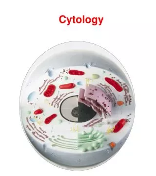
Cytology and Cytological Techniques Clinical Pathology
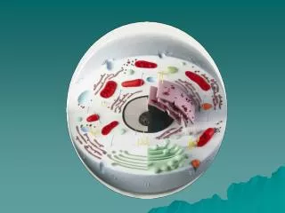
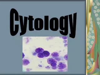
Cytology • The microscopic examination of cells. • Generally refers primarily to cells exfoliated from tissues, lesions, and internal organ/tumor cells. • A very valuable diagnostic tool. • Is inexpensive • Is quick and easy • Involves little or no risk to the patient
Cytology Continued • Must be able to identify normal cells from abnormal cells, and inflammatory from non-inflammatory cells • Disadvantage may be that some tumors do not exfoliate cells well and therefore may not provide and adequate sample to examine.
Cytologic Interpretation • May be able to diagnose • Identify the disease process • Help form a prognosis • May determine what diagnostic procedures should be performed next • May help with therapy options
Cytologic Techniques • Fine Needle Aspirate (FNA) • Fluid Aspiration- Thoracocentesis/Abdominocentesis • Solid mass imprinting • Vaginal wall technique • Cerebrospinal (CSF) Fluid Analysis • Synovial Fluid Analysis • Nasal Flush
General Collection Techniques • When possible prepare several smears • Use stained and unstained techniques • May use a variety of stains • Use clean, dry slides
Scrapings • Done on freshly cut surfaces • Scrap lesion/tissue with clean scalpel blade • Place material collected on a slide and spread • Advantage: May collect more cells • Disadvantage: More difficult to collect and only able to collect superficial lesions
Imprints • May be prepared from external lesions (ulcers) • May be prepared from tissues excised during surgery or necropsy. • Easy to collect • Disadvantage: May only collect few cells and may contain contamination
Solid Mass imprints • Cut mass in half • Blot dry • Need to remove blood/tissue fluid from surface • Use sterile gauze or other absorbent material • Excess blood/fluid inhibits cells from spreading and assuming normal size and shape • Touch the slide to the blotted surface • Stain
Fine Needle Aspirates • Preferred method of obtaining samples from masses. • Avoids superficial contamination • Very little risk to patient • Less complications to internal organs than core biopsy techniques • Implantation of malignant cells along the aspiration tract is extremely rare • Disadvantage: May not get a good sample because using just a small needle.
Fine Needle Aspirate • 2 techniques • Aspiration • Collect with 22-25 gauge needle • Use 3-12 ml syringe • Need slides • Non-aspiration
FNA Aspiration Technique • Hold mass/lymph node firmly • Introduce the needle with syringe attached into the mass • Apply strong negative pressure by withdrawing the plunger to about 2/3 -3/4 of the volume. • Do several times in same area or redirect needle. • Stop negative pressure and remove needle from mass • Remove needle from syringe and air is drawn up into syringe • Sample that is in hub of needle is expelled onto slide by rapidly depressing the plunger • Hold needle close to slide, if too far away will result in small droplets that dry rapidly before smear technique may be done.
FNA Non-Aspiration Technique • Works best for small masses that are difficult to aspirate. • Works well for highly vascular tissues • Using a needle only, move rapidly back and forth (stabbing motion). • Withdraw needle and place syringe with air to force onto slide.
Preparation of smears from aspirates • Squash prep method • Needle spread method • Blood smear method
Squash Preparation • With experience, can yield excellent cytologic smears • Aspirated material is placed on the center of the slide • A second slide is placed over the sample to form a cross. • Carefully slide apart from first slide (Put down on and pick up to move). • Do not place excessive downward pressure to the first slide because will cause distorted ruptured cells • The weight of the spreader slide is sufficient to adequately spread the cells.
Needle Spread Method • Spread aspirate on the slide with tip of needle. • Pull sample out into several projections (starfish appearance).
Blood Smear Technique • Use if material is thick or fluid • After material is expelled on slide, second slide is held at 30-40˚angle. • Second slide is pulled backward until it contacts the fluid • Rapidly move forward like a blood smear.
Common Problems with FNA • Few or no cells obtained • Some lesions do not exfoliate cells well. • The needle may miss the site of the lesion • Timid collection • Inadequate negative pressure • Blood contamination • Using too large needle gauge • Prolonged aspiration • Failure to blot if doing imprint
Common Problems with Preparation • Poorly prepared slides due to thick or high cell numbers • Allowing material to dry on slide before squash prep or other smear technique. • If a large amount of material is present, spread between two slides • May have to do 4-5 slides form the same site in order to get valuable diagnostic sample.
Staining Slides • Diff-quik, Wright’s, Geimsa • Papanicolau stains- • used in human Ob/gyn exams. Stains nucleus and nuclear material better. • New Methylene Blue stain • Air dry these slides, do not heat fix. • Use clean slides (make sure no lint on slide) • Stain immediately after air drying • Take care not to touch the surface of the slide or smear at any time.
Medical Terminology • Hypertrophy-an increase in cell size and/or functional activity in response to a stimulus. • Hyperplasia- increase in cell numbers, via increased mitotic activity, in response to a stimulus. • Neoplasia- increase in cell growth and multiplication that is not dependent on an external stimulus. • Metaplasia- a reversible process in which one mature cell type is replaced by another mature cell type (adaptive response to a stimulus)
Medical Terminology Continued • Dysplasia- reversible, irregular, atypical, proliferative cellular changes in response to irritation or inflammation. • Anaplasia- A lack of differentiation of tissue cells • Less differentiated cells in a tumor is more malignant • Chromatin pattern- the microscopic pattern of nuclear chromatin (the chromatin pattern coarsens as malignant potential increases)