Download
1 / 29
400 likes | 761 Views
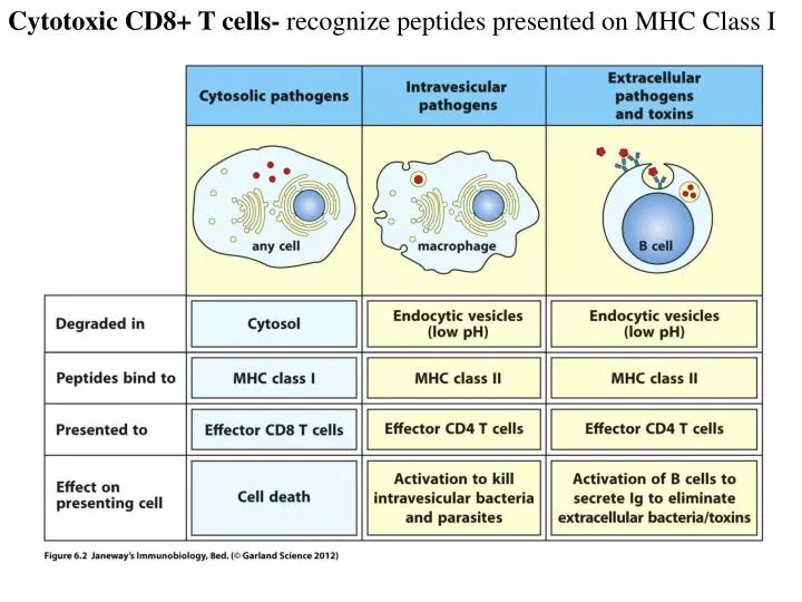
Cytotoxic CD8+ T cells- recognize peptides presented on MHC Class I. Upon recognition of peptide presented with MHCI on a target cell, CD8+ T cells kill the infected cell through release of cytotoxic granules.

E N D
Cytotoxic CD8+ T cells- recognize peptides presented on MHC Class I
Upon recognition of peptide presented with MHCI on a target cell, CD8+ T cells kill the infected cell through release of cytotoxic granules.
Following infection, CD8+ T cells expand, then contract to form a stable, homeostatic proliferating memory pool. Immunologic considerations for generating memory CD8 T cells through vaccination Noah S. Butler,Jeffrey C. Nolz,John T. Harty Cellular Microbiology ,Volume 13, Issue 7, pages 925–933, July 2011
Most current vaccines rely on neutralizing antibody induction, which may be ineffective at protecting against cell-mediated pathogens such as Malaria, TB, or HIV.
CD8+ T Cell Differentiation- Following infection, CD8+ T cells differentiate into a heterogeneous pool of cells with different memory potential. KLRG1 CD127 Short-lived Effector Cell (SLEC), KLRG-1+, CD127- Memory Precursor Effector Cell (MPEC), KLRG-1-, CD127+ Based on : Joshi et al (2008). Immunity, Sarkar et al (2008). JEM, Obar et al (2010) . PNAS, Obar & Lefrancois (2010) J Immunol
Factors controlling CD8+ T cell Differentiation- IL-12, IFN-, and IL-2 are known to drive terminal effector cell differentiation. T-bet and Blimp-1 are important transcription factors in these cells. Adapted from: Cui, W., Kaech, S. (2010). Generation of effector CD8+ T cells and their conversion to memory T cells. Immunol Reviews 236(1):151-166.
Mouse models for studying CD8+ T cells- Both ListeriaMonocytogenes and Vesicular Stomatitis Virus induce robust CD8+ T cell response and form stable memory pools. ListeriaMonocytogenes(LM) Vesicular Stomatitis Virus (VSV) Adapted from: Hamon, M., Bierne, H., Cossart, P. (2006) Listeriamonocytogenes: a multifaceted model. Nat Rev Micro 4(6):423-34. and:
E and Id proteins- Id proteins negatively regulate E proteins from binding DNA. It was previously shown that Id2 mediates CD8+ T cell immunity after infection. ** ** The function of E- and id proteins in lymphocyte development Isaac Engel & CornelisMurre Nature Reviews Immunology 1, 193-199 (December 2001)
The transcriptional regulators Id2 and Id3 control the formation of distinct memory CD8+ T cell subsets Cliff Y Yang,J Adam Best, Jamie Knell, Edward Yang, Alison D Sheridan, Adam K Jesionek, Haiyan S Li, Richard R Rivera, Kristin Camfield Lind, Louise M D'Cruz, Stephanie S Watowich, CornelisMurre& Ananda W Goldrath Nature Immunology Volume: 12, 1221–1229 (2011)
Supplemental Figure 1- Generation of Id2-YFP knockin reporter -Inserted YFP into Id2 locus -Also used a previously generated Id3 GFP reporter -When used as a heterozygote, no defects in lymphocyte development were observed (Id2Y/+ and Id3G/+) -Also made Id2Y/+ Id3G/+ OT-I mice, so all CD8+ T cells express a TCR that recognizes chicken ovalbumin (OVA)
Figure 1: Expression of Id2-YFP and Id3-GFP reporters in CD8+ T cells during infection identifies distinct effector populations. VSV-OVA LM-OVA -Transferred Id2Y/+ Id3G/+ CD45.1+ OT-I CD8+ T cells into CD45.2+ Bl/6 mice one day before infection with VSV-OVA
Figure 1: Expression of Id2-YFP and Id3-GFP reporters in CD8+ T cells during infection identifies distinct effector populations. VSV-OVA LM-OVA -Transferred Id2Y/+ Id3G/+ CD45.1+ OT-I CD8+ T cells into CD45.2+ Bl/6 mice one day before infection with VSV-OVA -Saw early Id3 down-regulation following infection with gradual re-expression at later timepoints (Day 14, 80) which correlates with memory formation
Figure 2: Expression of Id2-YFP and Id3-GFP during infection correlates with effector and memory precursor subsets. VSV-OVA
Figure 2: Expression of Id2-YFP and Id3-GFP during infection correlates with effector and memory precursor subsets. VSV-OVA -Id3hi cells have memory markers (CD127+, CD62Lhi) while Id3lo cells have the short-lived marker KLRG-1. Id3hi cells also make more cytokines??
Figure 3: The Id3-GFPhi effector CD8+ T cell population includes long-lived memory precursor cells before expression of KLRG-1 or CD127. -5 Days following infection, Id3 High and Low populations (KLRG-1 and CD127 negative) were sorted and transferred into infection-matched recipients.
Figure 3: The Id3-GFPhi effector CD8+ T cell population includes long-lived memory precursor cells before expression of KLRG-1 or CD127. +3 Days (Day 8 LM-OVA) +6 Days (Day 11 LM-OVA)
Figure 3: The Id3-GFPhi effector CD8+ T cell population includes long-lived memory precursor cells before expression of KLRG-1 or CD127. +3 Days (Day 8 LM-OVA) +6 Days (Day 11 LM-OVA) -At +3 Days, the Id3lo cells had more KLRG-1 expression than the Id3hi cells -At +6 Days, the Id3hi cells had more CD127 expression than the Id3lo cells -Proliferation of these two subsets was equal, so the Id3hi cells must survive better -After rechallenge of transferred cells, the Id3hi subset recalled preferentially
Figure 4: Early Id3 expression correlates with memory potential. -Id3hi cells correlate with genes up-regulated in CD127hi cells 1.5x -Id3hi cells correlate with genes up-regulated in memory cells -Id3lo cells correlate with genes up-regulated in effector cells
Figure 4: Early Id3 expression correlates with memory potential. -Transcripts associated with memory cells were up-regulated in Id3hi cells -Transcripts associated with terminal differentiation were down-regulated in Id3hi cells -All genes in green demonstrate putative E2A binding sites in their enhancer elements
Figure 5: Id3 deficiency results in the defective formation of long-lived memory CD8+ T cells. -Id3G/G mice= Id3 deficient -Mixed Id3G/G and WT OT-Is and transferred equal numbers into WT mice one day before VSV-OVA infection 80 days post VSV-OVA
Figure 5: Id3 deficiency results in the defective formation of long-lived memory CD8+ T cells. -Id3G/G mice= Id3 deficient -Mixed Id3G/G and WT OT-Is and transferred equal numbers into WT mice one day before VSV-OVA infection -Id3G/G and WT OT-Is had similar numbers and phenotype 7 days after infection -At 60 days post-infection, more WT OT-Is are present although the phenotype between the KO and WT are similar -80 days post-infection, a similar amount of BrdU incorporation is seen, but Id3G/G cells have higher cytokine production **The WT cells really have only twice as much memory generation** 80 days post VSV-OVA
Figure 6: Id2-deficient CD8+ T cells upregulate Id3 and do not generate short-lived effector-memory cells. 15 days post LM-OVA
Figure 6: Id2-deficient CD8+ T cells upregulate Id3 and do not generate short-lived effector-memory cells. 15 days post LM-OVA -Id2 deficiency results in Id3 up-regulation, but not visa versa -With Id2 deficiency and Id3 up-regulation, CD8 T cells are driven to a memory phenotype -Does Id2 directly regulate Id3?
Figure 7: Id3-GFP expression and Id2-YFP expression are inversely coregulated by cytokines. -In vitro treatment of Id2Y/+ Id3Y/+ OTI cells with OVA-peptide pulsed APCs with or without cytokines for 4 days
Figure 7: Id3-GFP expression and Id2-YFP expression are inversely coregulated by cytokines. -In vitro treatment of Id2Y/+ Id3Y/+ OTI cells with OVA-peptide pulsed APCs with or without cytokines for 4 days -Saw Id3 down-regulation with an Id2 up-regulation following treatment with IL-2, IL-12 and IL-21
Figure 7: Id3-GFP expression and Id2-YFP expression are inversely coregulated by cytokines. 15 days post LM-OVA
Figure 7: Id3-GFP expression and Id2-YFP expression are inversely coregulated by cytokines. 15 days post LM-OVA -STAT proteins signal downstream of many cytokines -STAT binding sites were found in the Id2 promoter and STAT4 and STAT5 were found enriched at these sites 7 days post infection with chromatin immunoprecipitation (ChIP) -In vivo, OT-I cells without exposure to IL-12 (IL-12 deficient host) had higher Id3 expression, again suggesting that IL-12 results in lower Id3
Conclusions: -Id3 expression is down-regulated following infection and slowly comes back on with time (like memory markers) -Sorted Id3-GFPhieffector cells “preferentially” gave rise to long-lived memory -The gene profile of Id3hi cells was similar to gene profiles of CD127hi memory precursors -Id3 deficient cells produced a smaller memory population than WT cells -Id2 deficiency resulted in the up-regulation of Id3 and progression to memory phenotype -IL-2, IL-12, and IL-21 (which drive CD8 T cells away from memory) caused up-regulation of Id2 and down-regulation of Id3 **Published back-to-back with another paper by Ji, Y. et al “Repression of the DNA-binding inhibitor Id3 by Blimp-1 limits the formation of memory CD8+ T cells**