Download
1 / 49
490 likes | 631 Views
DNA Deoxyribonucleic Acid. (a little background, more in cell biology; Chapter 16). Background. Genes are portions of DNA molecules that contain genetic information that controls protein synthesis. DNA is one of the nucleic acids and RNA is the other.

E N D
DNADeoxyribonucleic Acid (a little background, more in cell biology; Chapter 16)
Background Genes are portions of DNA molecules that contain genetic information that controls protein synthesis. DNA is one of the nucleic acids and RNA is the other. Each chromosome is made up of DNA and associated proteins. The DNA of each chromosome contains many genes.
Gene Expression The process whereby genes direct protein synthesis is called gene expression. DNA > RNA > protein DNA is transcribed into RNA RNA is translated into proteins DNA is also replicated (duplicated)
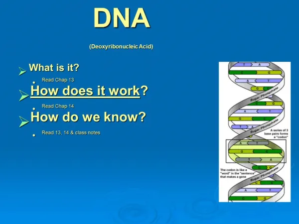
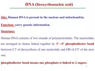
DNA structurePart I DNA is composed of subunits (nucleotides) each consisting of a 5Carbon sugar (deoxyribose), phosphate group, & nitrogenous bases Adenine (A), Thymine (T), Cytosine (C), and Guanine (G). Nucleotides are linked by sugar-phosphate bonds that basically form a backbone (like the sides of a ladder).
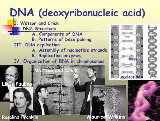
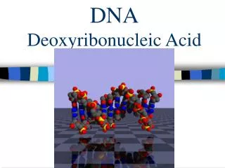
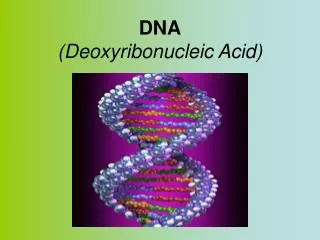
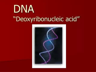
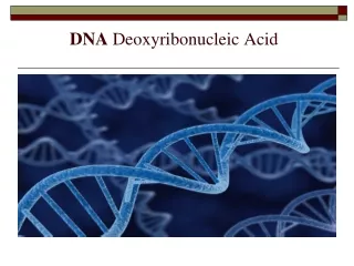
DNA structurePart II Base pairing between A and T and between C and G connects two strands (sides of ladder) together (like the steps of a ladder). The 3-D structure of DNA is a twisted spiral (or more commonly called a helix) consisting of two strand of DNA, thus it is called a double helix.
DNA Replication Part I In addition to serving as the code for proteins, DNA also undergoes replication. Replication is the duplication of DNA within a cell prior to cell division (occurring before mitosis & before meiosis I)
DNA Replication Part II The original strands of the double helix serve as a template for the new strands (replication is semi-conservative) 1. The double strand of DNA unwinds and unzips. 2. Then new DNA nucleotides base pair with those of the two original strands (C with G, and A with T) Enzymes are involved in replication (e.g., helicase and DNA polymerase)
Original – and New - T-A T-AT-A A-T A-TA-T C-G C-GC-G C-G C-GC-G T-A T-AT-A G-C G-CG-C G-C G-CG-C A-T A-TA-T T-A T-AT-A
Chromosome structure.Part I DNA (about 40%) + associated proteins (about 60%) = chromosomes Histones are the chromosomal proteins of eukaryotes
Chromosome structure.Part II Nucleosomes are bead-like particles containing DNA wound around clusters of histone proteins DNA can either be wound tightly (condensed) or relaxed (when replication or transcription are occurring; but in different ways)
Mitosis (Chapter 12)
Background information Part I Mitosis is part of the cell cycle and is the process whereby chromosomes divide followed by cell division (cytokinesis). It produces two identical daughter cells (these two cells have the same contents as the parent cell).
Background information Part II Meiosis is the two stage (two divisions; Meiosis I and Meiosis II) process whereby four (or in some cases only one) daughter cells receive only part of the contents of the parent cell. In particular, the daughter cells receive only half of the number of chromosomes. Meiosis is a special process that occurs in the formation of the sex cells or gametes in sexually reproducing organisms (animals and plants). However, replication occurs only before Meiosis I.
More Background In eukaryotes, replication results in the presence of two sets of each chromosome in the cell. These replicated chromosomes are most closely attached by a structure called a centromere and are known as sister chromatids.
In most higher animals and plants the chromosomes are paired You have 23 pairs of chromosomes, a total of 46: 23 from your mom & 23 from your dad. The members of these pairs are homologues. The two chromosomes of each pair carry genes controlling the same inherited characteristics. After replication there will be 46 x 2 = 92 total chromosomes present in a given human cell. Remember that the replicates will be physically attached at a centromere, so that it will still look like you have only 46 chromosomes In humans and other animals, there are two types of chromosomes: sex chromosomes autosomal chromosomes or autosomes are the other chromosomes
Background continued If an organism has two pairs of chromosomes, then this organism is said to be diploid (2n). If an organism or a cell (like our gametes) has only one set of unpaired chromosomes, then that organism or cell is said to be haploid (n). Some organisms are triploid, tetraploid (or just plain polyploid meaning many sets).
More Background The gametes (in human sperm and ova) are haploid (in humans n= 23 chromosomes) and thus when fertilization or syngamy occurs the diploid number is restored in the diploid zygote. Germ cells are those that undergo meiosis and somatic cells (non reproductive cells) are those that divide only by mitosis.
STAGES OF MITOSIS Prophase Metaphase Anaphase Telophase PMAT
Prophase The chromosomes shorten and thicken and become visible using a compound light microscope. The nuclear membrane breaks down In animals, the centrioles move to the opposite ends of the cell Spindle fibers appear
Metaphase The chromosomes line up at the equator of the cell (note the arrangement and compare it to Metaphase I of meiosis) The spindles are complete
Anaphase The centromeres divide The sister chromatids separate and are pulled toward the opposite poles
Telophase The sister chromatids have reached the opposite ends of the cell The nuclear membranes form around each set of chromosomes In most cases, the cytoplasm begins to divide
Cytokinesis the parent cell divides into two daughter cells (not a stage of mitosis)
MEIOSIS continued Like mitosis, meiosis can also be divided into stages, but it is more complex than mitosis. Meiosis involves two sets of stages or two PMATS.
STAGES OF MEIOSIS Meiosis I Prophase I Metaphase I Anaphase I Telophase I Cytokinesis Meiosis II Prophase II Metaphase II Anaphase II Telophase II Cytokinesis
Prophase I The chromosomes shorten and thicken and become visible using a compound light microscope The nuclear membrane breaks down In animals, the centrioles move to the opposite ends of the cell Spindle fibers appear The homologues pair up (remember these are not the same as sister chromatids which are already paired up). Crossing over occurs. This process involves the swapping of DNA between the homologues. It is an important source of variation.
Metaphase I The chromosomes line up at the equator of the cell, but the homologues not the sister chromatids are arranged toward the opposite ends of the cell. The spindles are attached to the homologues
Anaphase I The chromosomes begin to separate But, the homologues, not the sister chromatids are pulled apart
Telophase I Now the homologues are positioned at the opposite ends of the cell In some organisms, nuclear membranes reform Cytokinesis then occurs
Prophase II If the chromsomes lengthened, after cytokinesis they now shorten again
Metaphase II Spindles form and attach the chromosomes line up. Note the arrangement
Anaphase II Now the sister chromatids separate
Telophase II Now the sister chromatids of half of the original number of chromosomes are present in each of the haploid nuclei Nuclear membranes form Cytokinesis
Spermatogenesis In male animals, the gametes (sperm) are formed by a process called spermatogenesis Germ cells (spermatogonia or sperm mother cells) divide and some become primary spermatocytes (in the seminiferous tubules of the testes) Meiosis occurs producing four N daughter cells Immature gametes are called spermatids Spermatids mature into sperm with a head containing the nucleus, a tail (flagellum) and mitochondria In most males, sperm are produced continuously once puberty is reached
Oogenesis In female animals, the gametes (ova or eggs) are formed by a process called oogenesis Germ cells divide by mitosis and some become primary oocytes that divide (Meiosis I) into a secondary oocyte and a polar body. Thus there is an unequal distribution of the organelles and cytoplasm. This polar body may not undergo Meiosis II. The secondary oocyte divides (Meiosis II) into an ovum and another polar body (both N) In many animals, meiosis begins before birth and the females are born with primary oocytes arrested in meiosis I. This is related to chromosomal abnormalities in the children of older women.