Download
1 / 26
270 likes | 276 Views
Candi Jump, DO Asim Maqbool , MD Sudha Anupindi , MD Children’s Hospital of Philadelphia Reviewed by Melissa Jensen, MD of the Professional Education Committee. Pancreatitis. Case Presentation. 10 year old female presents to the ED 3 days of abdominal pain and vomiting

E N D
Candi Jump, DO AsimMaqbool, MD SudhaAnupindi, MD Children’s Hospital of Philadelphia Reviewed by Melissa Jensen, MD of the Professional Education Committee Pancreatitis
Case Presentation • 10 year old female presents to the ED • 3 days of abdominal pain and vomiting • History of 5 previous hospitalizations for similar symptoms
Case (Cont.) • FamHx: • Cholelithiasis in mother and MGM • Exam • Vital signs: normal • Abdomen: tenderness in epigastrium, non-distended • Labs • Amylase- 275 U/L (30-100 U/L) • Lipase- 600 U/L (15-130 U/L) • Ultrasound • Biliary sludge • Areas of calcification throughout pancreas
Presentation of Pancreatitis • Abdominal pain • Upper abdomen and radiating to the back • Vomiting • Abdominal distention • Tachycardia • Fever • Hypertension
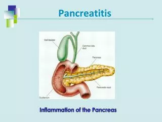
Acute Pancreatitis • Diagnostic criteria: presence of 2 of 3 features 1. Clinical symptoms: • Persistent abdominal pain 2. Elevation in serum amylase/lipase • >3 X upper limit of normal 3. Radiographic evidence of pancreatitis: • Pancreatic edema, calcifications, or pseudocyst • Further classified into • Interstitial • Necrotizing • Reversible, 10% of patients will have a recurrence
Chronic Pancreatitis • Irreversible • Diagnosis requires • Histologic or morphologic changes in the pancreas OR • Evidence of decreased digestive function (pancreatic insufficiency)
Etiology • Idiopathic • Systemic illness: • HUS, sepsis, sickle cell, DKA, Kawasaki • Biliary: • Anantomicabnormalitis (choledochal cyst), biliary obstruction (stones/sludge) • Anatomic: • Pancreatic divisum and annular pancreas • Drugs: • L-asparaginase, valproate, metronidazole, meracaptopurine, azathioprine, tetracycline • Genetics: • PRSS1, SPINK1, CFTR • Trauma • Metabolic: • Hyperlipidemia (hypertriglyceridemia), hypercalcemia, GSD, refeeding, Organic acidemias • Autoimmune pancreatitis • Postoperative
Biliary http://gallstoneflush.com/info.html
Anatomic Pancreatic Divisum http://radiographics.rsna.org/content/29/4/1003/F35.expansion.html
Pathophysiology • Traumatic disruption/obstruction of ducts • Trypsin lingers in tissue and activates • CFTR mutation • decreased ductular cell secretion with inspissated secretions and poor trypsin clearance, in those patient with pancreatic sufficiency • Hyperstimulation of Ca++ rise • premature activation of trypsinogen within acini • Cationic trypsinogen gene mutation (PRSS1) • trypsin cannot auto-inactivate • SPINK 1 mutation • activated trypsin within organ is unchecked • Hypertryglyceridemia • Mechanism unknown • Autoimmune
Diagnosis of Acute Pancreatitis • History • Symptoms • Medications, Trauma • Family history • Physical Exam • Abdominal tenderness • Jaundice • Turner’s or Cullen’s sign
Cullen’s and Turner’s Sign http://www.thelancet.com/journals/lancet/article/PIIS0140-6736%2808%2960993-9/fulltext
Labs • Amylase • Peaks at 48 hours • Elevated for 4 days • Falsely elevated: • SBO, mesenteric ischemia, tubo-ovarian disease, renal insufficiency • Lipase • Higher Specificity • Elevated 8-14 days longer than amylase • Bilirubin and liver function panel • Serum triglycerides and calcium • Consider genetic testing if recurrent pancreatitis
Imaging • Ultrasound • Always the first line, has limitations • Can detect: • Altered echogenicity • Dilated pancreatic and bile ducts • Gallstones and biliary sludge • Pancreatic calcifications • Choledochol cysts and cystic fluid collections • MRCP • Ductal malformations • ERCP • Cases of unexplained, recurrent, or prolonged pancreatitis • Suspicion of duct disruption or structural defect • Has therapeutic potential
MRCP and ERCP http://www.healthhype.com/gallbladder-tests-ultrasound-ct-hida-scan-ercp.html
Treatment • IV hydrations • Pain control • Monitor for complications
Fluid Management • Close monitoring of fluid balance • Consider losses from vomiting, 3rd spacing, NGT • Early fluid resuscitation may improve outcome and prevent pancreatic necrosis • No pediatric data, based on adult studies
Pain Management • Parenteral narcotics via PCA • All opiates may increase sphincter of Oddi pressure and worsen course • Meperidine • Morphine • Longer half-life • Fewer side effects
Nutritional Therapy • Pancreatic rest • Mainstay of therapy, still used in most institutions • Little evidence to support this approach • Adult data suggests early feeding: • Feeding initiated within 24 hours of admission after patient stabilized • No pancreatic rest required for mild acute pancreatitis • Shorter hospital stay for patients who were initiated on feeds compared to fasting patients
How and What to Feed? • Nasogastric Vs. Nasojejunal • Jejunal feeds • Less stimulation of exocrine pancrease • Bypasses stomach for those with delayed gastric emptying • Decompression of stomach • Diet • No evidence for use of low fat diet over regular diet
Medications • Antacids to prevent gastritis • Antibiotic therapy • Aimed at gram negative organisms from the gastrointestinal tract • Impinem, 3rd generation cephalosporins • Controversial
Surgical Treatments • Rarely needed, but can include: • Endoscopic sphincterectomy +/- stent placement • Partial pancreatic resection • Total pancreatectomywith Islet cell autotransplantation
Complications • Pancreatic pseudocysts (15%) • Pancreatic necrosis rare • Pancreatopleural fistulae • Pancreatic insufficiency • Common with chronic pancreatitis • Fat (steatorrhea) and protein malabsorption • Monitor fecal elastase as marker of pancreatic sufficiency • Diabetes (late complication) • Pain – chronic, severe
Back to the Patient… • MRCP • Consistent with US, no abnormalities of biliary system noted • ERCP • Small biliary stones in drainage from gallbladder • Genetic testing • PRSS1 mutation positive
Resources • Banks P.A., Freeman M.L: Practice guidelines in acute pancreatisis. Am J Gastroenterol 2006; 101:2379-2400. • Lowe M.E. Pancreatitis. In: Wyllie R, Hyams HJS, Kay M, eds. Pediatric Gastorintestinal and Liver Disease. 4th ed. Philadelphia, PA; 2011. • Marik P.E.: What is the best way to feed patients with pancreatitis? CurrOpinCrit Care 2009; 15:131-138. • O’Keefe S.J., Sharma S.: Nutrition support in severe acute pancreatitis. GastroenterolClin North Am 2007; 36:297-312. • Werlin S.L., Kugathasan S., Frautschy B.C.: Pancreatitis in children. J PediatrGastroenterolNutr 2003; 37:591-595.