Download
1 / 24
240 likes | 435 Views
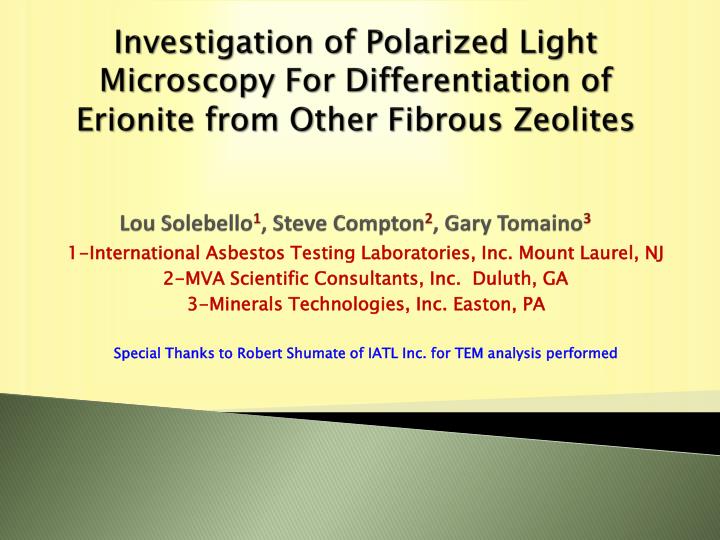
Investigation of Polarized Light Microscopy For Differentiation of Erionite from Other Fibrous Zeolites Lou Solebello 1 , Steve Compton 2 , Gary Tomaino 3. 1-International Asbestos Testing Laboratories, Inc. Mount Laurel, NJ 2-MVA Scientific Consultants, Inc. Duluth, GA

E N D
Investigation of Polarized Light Microscopy For Differentiation of Erionite from Other Fibrous ZeolitesLou Solebello1, Steve Compton2, Gary Tomaino3 1-International Asbestos Testing Laboratories, Inc. Mount Laurel, NJ 2-MVA Scientific Consultants, Inc. Duluth, GA 3-Minerals Technologies, Inc. Easton, PA Special Thanks to Robert Shumate of IATL Inc. for TEM analysis performed
Background Information • This investigation is part of a currently informal study performed by active members of the American Society for Testing Materials (ASTM). member interest is in characterization of erionite specimens and site specific samples containing erionite that may lead to development of standard test methods. Traditional environmental laboratory methods of analysis (XRD, SEM, TEM, PLM) are used for characterization. • Erionite is a naturally occurring, fibrous zeolite mineral with properties and health effects similar to asbestos. It usually is found in volcanic ash/tuffs that has been altered by weathering and ground water. Erionite is classified as an IARC Class I carcinogen based on epidemiology studies from exposure in the Cappadocia region of Turkey (IARC monographs 42:225-239, 1987). Erionite is not currently regulated by the U.S. Environmental Protection Agency (EPA) in the same manner as asbestos. • Cappadocia studies promoted increased awareness of exposure risk potential resulting from gravel pit excavation and disturbance of rock formations/soils during engineering activites that may contain zeolites. Identification of erionite occurrences and exposure risk has increased since the 1970’s Cappadocia study. • The North Dakota Department of Health, in cooperation with EPA, is currently conducting erionite exposure risk from Arikaree, Brule and Chadron geologic formations (Chalky Buttes, Little Badlands and Killdeer Mountain areas) in Slope, Stark and Dunn counties of North Dakota. • Procurement of reliable zeolite specimens can be challenging due psuedomorphism, difficulty in hand specimen identification without proper analytical confirmation, changes in mineral name classification and nomenclature.
Investigation Objectives • Initial focus on comparative characterization of verified erionite and other fibrous elongate zeolite specimens. Verified reference specimens from Rome Oregon, Cappadocia Turkey and Durkee Oregon are in the process of being characterized by ASTM members. A multi-analytical approach for characterization and identification of erionite is anticipated. Other fibrous/elongate zeolite and erionite specimens procured from various sources have been analyzed previously and reported on. Analytical methods commonly used in environmental and mineral R&D laboratories are and have been used for this study: 1. X-Ray Powder Diffraction (XRD)-Accepted definitive structural Identification/verification of zeolites. 2. Scanning Electron Microscopy (SEM) & Energy Dispersive X-Ray Analysis (EDX)- Morphology & cation analysis. 3. Transmission Electron Microscopy (TEM) & EDX-Morphology and cation analysis. Focus on 4. Polarized Light Microscopy (PLM) & Central Stop Dispersion Staining (CSDS) using a bench formulated High Dispersion (HD) 1.47 Refractive Index Liquid (RIL)-possible screening technique for differentiation of erionite from other fibrous/elongate zeolites.
Zeolite Conundrum • Identification of Zeolites: 1. Complex, naturally occurring framework aluminosilicate minerals (tectosilicate group). Characterized from other framework silicates, and differentiated from each other by the presence and number of water molecules. 2. The International Commission on New Minerals and Mineral Names (CNMMN) recognized general zeolite formula: (Ca,Na2,K2,Ba,Sr,Mg,Cs2,Li2)a[AlaSin-a02n] •xH2O. 3. There are 82 CNMMN recognized mineral species (13 compositional series) and over 150 synthesized zeolites. 4. Identification of zeolite species can be challenging due to similarities in physiochemical properties.
XRD Identification Summary of Specimens Analyzed to Date SpecimenXRD Validation Karain, Cappadocia Turkey Erionite Erionite/Qtz/Anor Durkee OR Erionite Erionite Rome OR Erionite Erionite/chabazite/qtz/Anor Chase Creek, Erionite Offretite (sold as erionite) Beech Creek, Erionite-Ca Chabazite and Thomsonite Mordenite, Goble Cty Mordenite Thomsonite, Sagasen Natrolite Natrolite , Lane Cty Natrolite Mesolite, Skookumchuck Mesolite
Example XRD Patterns For Validation Karain, Cappadocia Turkey Erionite Rome Oregon Erionite Chase Creek Offretite (sold as Erionite) Durkee Oregon Erionite
XRD Observations • ICDD card matches for Rome Oregon and Cappadocia (Karain) Turkey erionite same (04-010-5070 & 000-012-0275). Peak intensity/shape/symmetry suggests high crystallinity. Suggests Rome & Karain have similar chemistry. PLM examination consistent. • ICDD card match for Durkee Oregon slightly different compared to Rome and Karain (no 000-012-0275). Suggests different chemistry. Peaks suggest poor cyrstallinity, possibly differences in structure compared to Rome & Karain. • Accessory mineral XRD identification for Rome and Karain same (quartz, chabazite, anorthite). Hand specimen and PLM examination consistent.
Durkee Oregon Erionite TEM Tem Image 40,000X EDX Data
Rome Oregon Erionite TEM EDX Data TEM Image 12,000X
Karain (Cappadocia) Erionite TEM TEM Image 5,000X EDX Data
Chase Creek Offretite (Sold as Erionite) TEM TEM Image 8,000X EDX Data Compositional Variation on Ca, Mg, K, Na along fiber lengths. One end K+Na>Mg+Ca, other end reversed. Suggests erionite intergrowths
Select Area Electron Diffraction (SAED) Durkee Erionite Pattern not indexed at date of presentation, further work to be performed in future. Limited SAED suggests hexagonal pattern, diffuse (not sharp) spots expected for erionite.
EDX Elemental Ratios---Patterns? TEM Observations 1-Rome, Karain and Durkee Erionite specimens have similar morphology by TEM, similar to Offretite (Chase Creek). 2-Chase Creek specimen EDX analysis indicates distinctly different chemical composition at ends of fibers. This suggests offretite with intergrowths of erionite (not detectable by XRD). Na+K>Mg+Ca domains suggest erionite, Mg+Ca>Na+K domains suggest offretite. 3- Erionite specimens examined have similar EDX spectra, quantitative elemental analysis indicates variation of cation content (expected) for six fibers analyzed for each specimen. 4-Comparison of elemental ratios (table above) suggests that Si:Al, Si:K and Al:K may be useful for differentiating erionite from other fibrous zeolites on the basis of the limited data acquired at time of this presentation. Further work on other erionite and fibrous zeolite specimens required.
Erionite Differentiation Using 1.47 CSDS Concept • Erionite Identification Screen by PLM-Process of Elimination: 1. CSDS is an optical microscopy contrast enhancement technique for measuring RI that is more sensitive than the Becke-line method. It is an established asbestos minerals identification technique using 1.550 (chrysotile), 1.605 (actinolite,tremolite, anthophyllite) and 1.680 (amosite, crocidolite). Magenta blue CSD of a particle indicates a “ RI match” with a liquid, blue CSDS indicates the RI of the particle is less than the liquid, and gold CSDS indicates. the RI of the particle is greater than the liquid. 2. A limited number ( approximately 15) of the 82 naturally occurring zeolite species have documented fibrous and/or elongate particle morphology. 2. The range of index of refraction values for fibrous elongate zeolites: 1.45-1.52, most appear to be above 1.47. Reported/documented range of refractive index values for erionite: 1.455-1.485, depending on major cation (K, Ca, Na) prevalence. 3. 1.48, 1.49 and 1.47 HD RIL’s were made from1.550 Series E and triacetin for assessing the possibility of differentiating erionite from other fibrous zeolites using CSDS combined with sign of elongation, extinction angle, morphology and birefringence for erionite PLM/CSDS screening protocol similar in principle to what is used for asbestos identification.
Consider PLM Method Limitations • Particle Size: 1μm width, 5 μm length. PLM method is not appropriate for detection or quantification of “respirable” erionite fibers, which requires SEM and/or TEM assessment of exposure potential. • PLM is not the accepted definitive means for structural identification of zeolites. (XRD) is required, but has limited detection limit capabilities based on current technology, and is not without erionite interferences in complex mineral mixtures such as soils and rock. • CSDS colors can be subdued at 100X magnification due to low birefringence of erionite particularly with fibers approaching resolution limits. Commercial 200X and 400X CSDS objectives are limited, can be fabricated, but require significantly more illumination than standard PLM microscope configurations in order to observe CSDS colors. • The PLM screening technique may require significantly more knowledge, experience and resources than what may be available to many analysts performing traditional environmental PLM/CSDS analyses (asbestos).
Confirmation of Liquid Refractive Index: 1.47 Liquid Example
Durkee Oregon “Wooly” Erionite Magenta CSDS parallel to fiber bundle length indicates match: RI=1.47. Blue CSDS perpendicular to fiber bundle length indicates RI of erionite is slightly less than 1.47. Morphology is similar to polyfilamentous morphology characteristic of chrysotile asbestos.
Karain, Cappadocia Turkey Erionite 1.47 CSDS Blue CSDS parallel and perpendicular indicates RI’s are slightly less than 1.47. Indicates compositionally different compared to Durkee Oregon erionite. Morphology is similar to tremolite-actinolite asbestos habit, and discernibly different from wooly Durkee erionite.
Rome Oregon Erionite 1.47 CSDS Blue CSDS colors parallel and perpendicular indicates RI is slightly less than 1.47. RI’s and morphology consistent compared to Karain, different compared to Durkee.
Rome Oregon Erionite 1.47 CSDS Digital magnification of acquired image accentuates CSDS better than can be acquired using 200X or 400X CSDS objective, confirming RI’ are slightly less than 1.47.
Chase Creek Offretite 1.47 CSDS Gold CSDS indicates RI is greater than 1.47. Blue magenta CSDS indicates RI is very slightly less than 1.47. Light blue CSDS indicates RI is less than 1.47. Distinctly different hexagonal prism morphology compared to needle-like Rome/Karain and polyfilamentous Durkee erionite. Difference in CSDS along fiber length indicates compositional variation, possibly structural variation (possible erionite intergrowth). Consistent with TEM EDX analysis.
Goble Creek Oregon Mordenite 1.47 CSDS Magenta blue CSD perpendicular to fiber length indicates RI is close to 1.47. Blue CSD parallel to fiber length indicates RI is less than liquid. Differences in CSDS compared to erionite specimens examined. Mordenite can be further differentiated from erionite and offretite on basis of sign of elongation. Mordenite (-), erionite (+), offretite (+/-).
1.48 CSDS and Two Other Zeolites Example of further Differentiation Skookumchuck Mesolite: “Anomalous” CSDS and morphologically different from erionite. Lane Cty Oregon Natrolite: 1.48 CSD different compared to erionite in 1.48 (light blue). Morphologically similar. 1.48 HD RI can further facilitate differentiation of fibrous/elongate zeolites
Summation • Rome Oregon and Cappadocia (Karain) Turkey erionite exhibit similar morphologies (TEM,PLM), chemistry (EDX) and structure (XRD). Suggests Rome Oregon erionite would serve as a suitable North American reference specimen for comparison to Cappadocia erionite upon which epidemiology studies for IARC classification is based. • TEM analysis suggests similar EDX patterns with variable chemistry as expected. Si:Al, Si:K and AL:K appear to suggest consistency and may be useful for differentiation of erionite from other fibrous zeolites. Further work is required to determine if the observed patterns are consistent for erionite and different for other fibrous zeolites. SAED analysis indicates indexable patterns obtainable for potential structural identification. Further work is required. • Differentiation of erionite from other fibrous/elongate zeolites by 1.47 PLM CSDS comparison as a screening technique shows promise. If consistency in future analysis is demonstrated, the technique may be used in conjunction with XRD, SEM and TEM analysis of specimens for erionite content characterization. • Further CSDS analysis combined with comparison of morphology and optical attributes (sign of elongation, birefringence) on more erionite & other fibrous/elongate zeolite specimens is required to demonstrate consistency in observations made to date on the limited number of specimens examined.