Download
1 / 74
740 likes | 864 Views
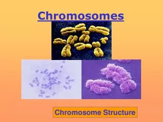
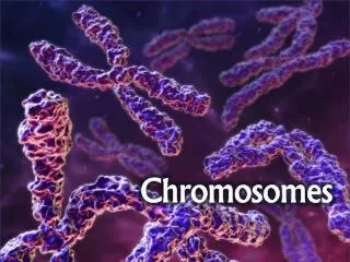
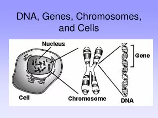
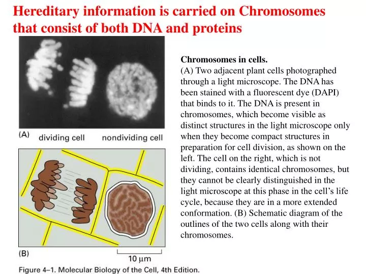
Hereditary information is carried on Chromosomes that consist of both DNA and proteins. Chromosomes in cells.

E N D
Hereditary information is carried on Chromosomes that consist of both DNA and proteins Chromosomes in cells. (A) Two adjacent plant cells photographed through a light microscope. The DNA has been stained with a fluorescent dye (DAPI) that binds to it. The DNA is present in chromosomes, which become visible as distinct structures in the light microscope only when they become compact structures in preparation for cell division, as shown on the left. The cell on the right, which is not dividing, contains identical chromosomes, but they cannot be clearly distinguished in the light microscope at this phase in the cell’s life cycle, because they are in a more extended conformation. (B) Schematic diagram of the outlines of the two cells along with their chromosomes.
Chromosomal DNA and its packaging in the chromatin fiber Eucaryotic DNA is enclosed in cell nucleus, and is packaged into a set of chromosomes
Chromosome Structure • Human DNA’s total length is ~2 meters! • This must be packaged into a nucleus that is about 5 micrometers in diameter • This represents a compression of more than 100,000x • It is made possible by wrapping the DNA around protein spools called nucleosomes and then packing these in helical filaments
GENOME SEQUENCE AND CHROMOSOME DIVERSITY Chromosomes can be circular or linear
The E. coli genome is composed almost entirely of genes More complex organisms have decreased gene density
Genes make up only a small portion of the eukaryotic chromosomal DNA Representation of the nucleotide sequence content of the human genome. LINES, SINES, retroviral-like elements, and DNA-only transposons are all mobile genetic elements that have multiplied in our genome by replicating themselves and inserting the new copies in different positions. Simple sequence repeats are short nucleotide sequences (less than 14 nucleotide pairs) that are repeated again and again for long stretches. Segmental duplications are large blocks of the genome (1000–200,000 nucleotide pairs) that are present at two or more locations in the genome. Over half of the unique sequence consists of genes and the remainder is probably regulatory DNA. Most of the DNA present in heterochromatin, a specialized type of chromatin that contains relatively few genes, has not yet been sequenced
The majority of human intergenetic sequences are composed of repetitive DNA Microsatellite DNA (simple sequence repeats) Genome-wide repeats (transposable elements) LINES : (Long Interspersed Sequences)
CHROMOSOME DUPLICATION AND SEGREGATION Eukaryotic chromosomes require centromeres, telomeres, and origin of replication to be maintained during cell division
Centromere size and composition vary dramatically among different organisms
Eukaryotic chromosome duplication and segregation occur in separate phases of the cell cycle
Sister-chromatid cohesion and chromosome condensation are mediated by SMC (structural maintenance of chromosome) proteins
During gap phases, cells prepare for the next cell cycle stage and check that the previous stage is completed correctly
Different levels of chromosome structure can be observed by microscopy
THE NUCLEOSOME DNA Molecules Are Highly Condensed in Chromosomes Nucleosomes Are the Basic Unit of Eukaryotic Chromosome Structure
Structural organization of the nucleosome. A nucleosome contains a protein core made of eight histone molecules. As indicated, the nucleosome core particle is released from chromatin by digestion of the linker DNA with a nuclease, an enzyme that breaks down DNA. (The nuclease can degrade the exposed linker DNA but cannot attack the DNA wound tightly around the nucleosome core.) After dissociation of the isolated nucleosome into its protein core and DNA, the length of the DNA that was wound around the core can be determined. This length of 146 nucleotide pairs is sufficient to wrap 1.65 times around the histone core.
Micrococcal nuclease and the DNA associated with the nucleosome
Nucleosomes as seen in the electron microscope. (A) Chromatin isolated directly from an interphase nucleus appears in the electron microscope as a thread 30 nm thick. (B) This electron micrograph shows a length of chromatin that has been experimentally unpacked, or decondensed, after isolation to show the nucleosomes.
Histones are small, positive charged proteins Core histones share a common structural fold
Amino-terminal tails of the core histones are accessible to proteases
The atomic structure of the nucleosome Histones bind characteristic regions of DNA within the nucleosome
Interactions of the histones with nucleosomal DNA H2A-H2B bind 30 bp of DNA on one side of nucleosome H3-H4 bind the middle and the ends of DNA
Many DNA sequence-independent contacts mediate the interaction between the core histones and DNA
The histone –N-terminal tails stabilize DNA wrapping around the octamer
Wrapping of the DNA around the histone core store negative superhelicity Removal of nucleosomes not only allows access to the DNA, but also facilitates DNA unwinding of nearby DNA Sequences
HIGHER-ORDER CHROMATIN STRUCTURE Heterochromatin is highly organized and unusually resistant to gene expression Heterochromatin: highly condensed higher-order structure forms that result in a barrier to gene expression Euchromatin:the nucleosomes are found to be in much less organized assemblies
Nucleosome arrays can form more complex structures; the 30-nm fiber
The histone tails are required for the formation of the 30- nm fiber. (Left): The approximate exit points of the eight histone tails, four from each histone subunit, that extend from each nucleosome. In the high-resolution structure of the nucleosomethe tails are largely unstructured, suggesting that they are highly flexible. (Right): A speculative model showing how the histone tails may help to pack nucleosomes together into the 30-nm fiber. This model is based on (1) experimental evidence that histone tails aid in the formation of the 30-nm fiber, (2) the x- ray crystal structure of the nucleosome, which showed that the tails of one nucleosome contact the histone core of an adjacent nucleosome in the crystal lattice, and (3) evidence that the histone tails interact with DNA.
Further compaction of DNA involves large loops of nucleosomal DNA