Download
1 / 41
410 likes | 556 Views
Announcements. Artemia reports due today: please put on front desk. Paper today: Wongprasert et al. 2003 (Katie leads) Paper for next week: Jud et al. 2007 (Molly leads) Start computers, open lecture PP for simulations again.

E N D
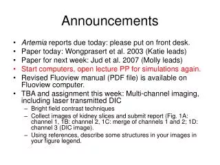
Announcements • Artemia reports due today: please put on front desk. • Paper today: Wongprasert et al. 2003 (Katie leads) • Paper for next week: Jud et al. 2007 (Molly leads) • Start computers, open lecture PP for simulations again. • Revised Fluoview manual (PDF file) is available on Fluoview computer. • TBA and assignment this week: Multi-channel imaging, including laser transmitted DIC • Bright field contrast techniques • Collect images of kidney slices and submit report (Fig. 1A: channel 1, 1B: channel 2, 1C: merge of channels 1 and 2; 1D: channel 3 (DIC image). • Using references, describe some structures in your images in your figure legend.
Paper Discussion Schedule • Today, Jan. 22 (Hertzler): Zucker 2006 • Jan. 29 (Katie): Wongprasert et al. 2003 • Feb. 5 (Molly): Jud et al. 2007 • Feb. 12 (Becky): Anders 1988 • Feb. 19 (Rachel): Tan et al. 2005 • Feb. 26 (Ellen): • March 12 (Emily) • March 19 (Amy) • March 26 (Amanda) • April 2 (Andrea) • April 9 (Brittaney) • April 16 (Lauren) • April 23 (Joe and Molly):
Outline: Contrast Enhancement, Confocal Hardware • Resolution and sampling frequency: XY and Z • Kohler Illumination and microscope setup • Contrast Enhancement • Brightfield • Interference of light • Phase contrast • Polarization • DIC • Components of LSCM • Scan Head • Lasers • Light Detectors • Week 4 TBA
Nyquist Sampling Theorem: XY • Hibbs, p. 126: “When a continuous, analogue image is digitised, the information content of the signal will be retained only if the diameter of the area represented by each pixel is 2.3x smaller than the optical resolution limit of the microscope.” • So an objective with a theoretical resolution of 0.2 μm requires a pixel size of 0.08 μm. • How do you determine the pixel size (sampling frequency)? • Measure it with your scale bar at different zoom factors: From: Pawley, 2006. Handbook of Biological Confocal Microscopy. Springer: New York.
Nyquist sampling of an image of two points separated by the Rayleigh resolution(Pawley 2006) Sampling interval = d/2.3 From: Pawley, 2006. Handbook of Biological Confocal Microscopy. Springer: New York.
5. Axial Resolution (Z or raxial) • Minimum distance between the 3D diffraction patterns (PSFs) of two points along the Z axis that can still be seen as two. • From: Pawley, 2006. Handbook of Biological Confocal Microscopy. Springer: New York.
5. Axial Resolution (Z or raxial) • So with η = 1.5 for methyl salicylate:
Ideal step size (higher Z resolution, e.g. NA=1.4) Ideal step sizes Ideal step size (lower Z resolution, e.g. NA=0.7) Undersampled (lower Z resolution, e.g. NA=0.7)
XY and Z resolutions (μm), XY Zoom andZ step sizes (1024 X 1024 box size) *Nyquist sample frequency of 2.3 +Nyquist sample frequency of 3.0
B. Kohler Illumination • Purpose: Bright, even illumination without illuminating unnecessary areas or excess flare. Steps • Focus on the sample. • Close field diaphragm until it can be seen, focus and center the condenser. • Open field diaphragm until it disappears from view.
Upright Scope Epi- illumination Source Brightfield Source
Inverted Microscope Brightfield Source Epi- illumination Source
C. Enhancing contrast in LMFibroblast in Culture: Four Types of Light Microscopy Bright-Field Phase-Contrast Differential Interference Contrast Dark-Field
1. Bright-field microscopy • Is the simplest, but object must be colored to be seen. Histological staining usually requires killing the sample. • Staining utilizes absorption; e.g. red stain absorbs green and blue light, passing only red light. The specimen is now an amplitude object, where contrast is seen by reducing the amplitude of certain wavelengths of light. • Microscopy of living cells, which are usually transparent, are limited by contrast, or the difference between light and dark. • How can we see them without staining them? • By exploiting the fact that samples are phase objects, which slow light down relative to other parts of specimen or to background.
3. Phase-Contrast Microscopy • Annular Ring in Phase Condenser focuses cone of light onto sample. • Specimen light is shifted -1/4 wavelength • In Phase ring of Objective: • Direct light (background) passes through thin, dark part. • Diffracted light (specimen) passes through thick, light part, shifted -1/4 wavelength. • Specimen light shifted by ½ wavelength total. • Rings must be aligned to get phase effect.
Alignment of phase rings • Jave tutorial: http://micro.magnet.fsu.edu/primer/java/phasecontrast/phasemicroscope/index.html ALIGNED
λ/2 Phase Contrast Microscope • Surround wave (red) is undiffracted light that passes around and through the sample. • Diffracted wave (blue) interacts with sample, is retarded by ¼ wavelength relative to S wave. • Particle wave (green) results from interference between S and D waves. Amplitude difference between S and P determines the level of contrast • PC scope shifts diffracted beam from specimen an additional ¼ wavelength to ½ λ, creating maximal destructive interference between S and D. • Causes decrease in amplitude (brightness) in P, which can be seen against brighter background. Object dimmer Background bright Object brighter Background dimmer
Limitations of Phase Contrast • Phase images are usually surrounded by halos around the outlines of details. Such halos are optical artifacts, which sometimes obscure the boundaries of details. • The phase annuli do limit the working numerical aperture of the optical system to a certain degree, thus reducing resolution. • 20X PlanApo 0.7 NA compared with 20X Phase 0.4 NA. • Phase contrast does not work well with thick specimens because shifts in phase occur from areas slightly below or slightly above the plane that is in focus. Such phase shifts confuse the image and distort image detail.
Useful for crystalline materials or oriented structures in biological materials, e.g. Mitotic spindle fibers Microfilament bundles Striated muscle fibers These structures are said to be birefringent (having double refraction), meaning that they have at least two refractive indices. 4. Polarization Microscopy Birefringent skeleton in sea urchin larva
Polarization of Light AKA Analyzer Note: laser light is already polarized
Polarizing Sunglasses • Human eye can’t detect difference in randomly oriented versus polarized light. • When polarizing sunglasses filter out parallel waves, eye detects less glare, lower amplitude.
Isotropic versus anisotropic materials Light slowed equally vibrating in any direction. Light slowed less when vibrating N-S n1 n1 n2 n1 Light slowed more when vibrating E-W n1 Isotropic: Glass, salt Anisotropic (birefringent – with two Refractive indices): Sugar, muscle, gout crystals
Birefringence • First clue to explanation of polarization came from observation of calcite crystals by Erasmus Bartholin in 1669. • http://www.microscopy.fsu.edu/primer/java/polarizedlight/icelandspar/index.html • One of the light rays emerging from a birefringent crystal is termed the ordinary ray, while the other is called the extraordinary ray. Split, both Polarized; perpendicular Incident ray oblique to optical axis of crystal
Incident light perpendicular to optical axis of specimen: same trajectory, different path length causes interference when recombined.
Birefringent samples oriented 45o to crossed polarizers are maximally bright • Java tutorial: http://micro.magnet.fsu.edu/primer/java/polarizedlight/crystal/index.html
5. Differential Interference Contrast • Also called Nomarski optics; uses plane polarized light. • Similar to Phase Contrast in that light from low contrast sample is caused to interfere destructively to produce amplitude changes. • Produces contrast where changes in thickness, slope, or refractive index occur in cell, especially along edges, to give a pseudo three dimensional appearance.
Cheek cells Filamentous alga Red blood cells DIC images have no halos.
DIC Pathway:Components • Light from lamp passes through Polarizer, is separated into O and E waves by Wollaston Prism (specific to each lens), then to Condenser. • Phase specimen creates different optical path lengths for O and E, shifting their phase. • After passing through specimen, light passes through Objective, and is recombined, resulting in interference, by second Wollaston Prism, then to Analyzer (second Polarizer) to Eyepiece.
Effect of Bias Retardation in Analyzer • Controls how O and E waves are recombined. • Affects brightness, contrast, and color (optical staining) of specimen. • Java tutorial: http://micro.magnet.fsu.edu/primer/java/dic/lightpaths/index.html
Advantages, Disadvantages of DIC • Advantages: • Uses full NA of the lens, achieving optimal resolution and some optical sectioning ability. • Provides optical color staining. • No phase halos as with phase contrast. • Main Disadvantage: • Tissue culture plastic or birefringent sample features can produce confusing effects.
1. Fluoview 300 Scan Head Anatomy 2 1 10 4 3 9 5 7 8 Beam splitter 6
2. Lasers available for Olympus Fluoview Confocal Microscopes • Blue argon-ion (488 nanometer) laser (WE HAVE) • Multi-line argon-ion (457, 488, and 514 nanometers) laser • Green helium-neon (543 nanometer) laser (WE HAVE) • Red helium-neon (633 nanometer) laser (WE MAY GET) • Yellow krypton-ion (568 nanometer) laser • Blue-violet helium-cadmium (442 nanometer) laser • Violet and blue-violet diode (405 and 440 nanometer) lasers • Ultraviolet argon-ion (351 nanometer) laser • Infrared (750 nanometer) laser
Adjusting Offset and PMT/Gain to maximize range of grey levels collected Pixel intensity 256 (4047) 0 Figure 5(a) illustrates the raw confocal image along with the signal from the photomultiplier. After first applying a negative offset voltage to the photomultiplier, the signal and image appear in Figure 5(b). Note that as the signal is shifted to lower intensity values, the image becomes darker (upper frame in Figure 5(b)). When the gain is next adjusted to the full intensity range (Figure 5(c)), the image exhibits a significant amount of detail with good contrast and high resolution.
Note on Building stereo images • Stereo Factor: Sets the deviation between the left and right eyes when building a pair of stereo 3D images or a 3D image to be viewed through color (red/green) eyeglasses. • You can change this. • Z Stretch Factor: Provides each section with a feeling of thickness. Usually this value does not need to be changed.
Week 4 TBA • Kohler illumination • Phase contrast • Polarization • DIC • Laser transmitted DIC • Assignment: Collect multi-channel fluorescence and laser transmitted images of prepared kidney slide, save, submit report as before.