Download
1 / 26
270 likes | 387 Views
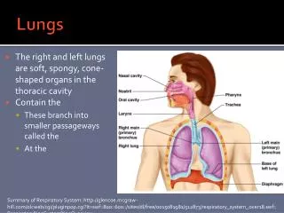
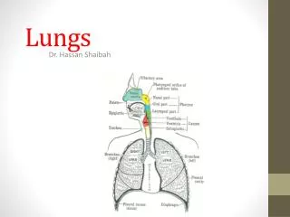
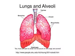
Airways and Lungs. Sanjaya Adikari Department of Anatomy. Nasal Cavity Nasopharynx (Oropharynx). Upper respiratory tract. Larynx Trachea Bronchi ………. Lower respiratory tract. Trachea Primary bronchi Secondary bronchi Tertiary bronchi Bronchioles Terminal bronchioles

E N D
Airways and Lungs Sanjaya Adikari Department of Anatomy
Nasal Cavity Nasopharynx (Oropharynx) Upper respiratory tract Larynx Trachea Bronchi ………. Lower respiratory tract
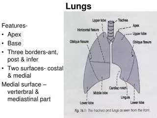
Trachea Primary bronchi Secondary bronchi Tertiary bronchi Bronchioles Terminal bronchioles Respiratory bronchioles Alveolar ducts Alveolar sacs Alveoli L & R bronchi Lobar bronchi Segmental bronchi
Nasal cavity • Hair at the entrance to trap bigger particles • Mucous and serous glands to produce secretions to trap smaller particles and moisturize the air • Highly vascularto increase air temperature • Folds on the nasal walls to increase the surface area • Pseudostratified columnar ciliated epithelium which traps and moves up the dust particles
General histological arrangement of the airway • Mucosa • Smooth muscle layer • Submucosa • Cartilage layer • Adventitia
Changes in the arrangement • PSCC epithelium in large air ways become simple cuboidal nonciliated epithelium in small air ways • Frequently seen goblet cells become less frequent and totally absent in terminal bronchioles • The layer of smooth muscles becomes thicker as it goes down and becomes maximum at terminal bronchioles • Serous and mucous glands in submucosal connective tissue becomes less numerous and absent beyond tertiary bronchi • Cartilage parts smaller in small airways
Trachea • Pseudostratified columnar ciliated epithelium • Smooth muscle layeralmost absent • Numerous serous mucous glandsin the submucosa • 'C' shaped hyaline cartilage
Primary and secondary bronchi • Less taller respiratory epithelium • Smooth muscle layerdiscontinuous • Fewer serous and mucous glands • Few hyaline cartilage plates rather than 'C' shaped ones
Tertiary bronchi • Tall simple columnar ciliatedepithelium • Smooth muscle layercontinuous • Very fewer serous and mucous glands • Few small irregular hyaline cartilage plates
Bronchioles • Airways < 1 mm in diameter • Simple columnar ciliatedepithelium • Smooth muscle layercontinuousand prominent • Serous and mucous glandsabsent • Cartilage plates absent
Terminal bronchioles • Structure is as same as the other bronchioles • End of the purely conducting portion of the airway Respiratory bronchioles • Walls contain small number of single alveoli • Ciliated cuboidal epithelium. No goblet cells
Alveolar ducts • Numerous alveolar sacs open into these • Simple cuboidal nonciliatedepithelium
Alveolar wall • Type I pneumocytes (The squamous cells) • Forms part of the gaseous diffusion barrier • Type II pneumocytes • Produce surfactant • Connective tissue • Reticulin, collagen and elastic fibres with fibroblasts. • Macrophages. • Blood vessels, mainly capillaries
Respiratory barrier/Gaseous diffusion barrier Type I pneumocyte Common BM Capillary endothelial cell
Alveolar pores • Equalize the pressure between the alveoli • Collateral circulation of air when the bronchiole is obstructed • Responsible for easy spread of infection eg. Lobar pneumonia