Download

1 / 51
510 likes | 520 Views
Pulmonary Function Testing The Basics of Interpretation. Jennifer Hale, M.D. Valley Baptist Family Practice Residency. Objectives. Identify the components of PFTs Describe the indications Develop a stepwise approach to interpretation Recognize common patterns

E N D
Pulmonary Function TestingThe Basics of Interpretation Jennifer Hale, M.D. Valley Baptist Family Practice Residency
Objectives • Identify the components of PFTs • Describe the indications • Develop a stepwise approach to interpretation • Recognize common patterns • Apply this information to patient care
Pulmonary Function TestingJennifer Hale, M.D. Which of the following is used to follow disease severity in COPD patients? • Total lung capacity (TLC) • Degree of responsiveness to bronchodilators • Forced vital capacity (FVC) • Forced expiratory volume in 1 second e. Diffusing capacity (DLCO)
Pulmonary Function TestingJennifer Hale, M.D. A 36yo WF, non-smoker, presents to your office for follow-up of ‘recurrent bronchitis.’ You suspect asthma and decide to order spirometry. Which of the following would you include in your prescription for testing? • Diffusing Capacity (DLCO) • If no obstruction present, add trial of bronchodilator • If no obstruction present, perform methacholine challenge • Flow volume loop • b and c
Pulmonary Function TestingJennifer Hale, M.D. A 68yo HM is admitted to the ICU with acute respiratory distress. A CXR obtained in the ED demonstrates bilateral pulmonary infiltrates, and his DLCO is elevated. What is the most likely diagnosis? • Pulmonary edema • Aspiration pneumonitis • Pulmonary emboli • Alveolar hemorrhage e. Interstitial lung disease
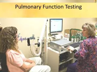
The Purpose Provide quantifiable, reproducible measurement of lung function
Description • Spirometry • Flow Volume Loop • Bronchodilator response • Lung volumes • Diffusion capacity (DLCO) • Bronchoprovocation testing • Maximum respiratory pressures • Simple and complex cardiopulmonary exercise testing
Indications — Diagnosis • Evaluation of signs and symptoms - SOB, exertional dyspnea, chronic cough • Screening at-risk populations • Monitoring pulmonary drug toxicity • Abnormal study - CXR, EKG, ABG, hemoglobin • Preoperative assessment
Indications — Diagnosis • Evaluation of signs and symptoms - SOB, exertional dyspnea, chronic cough • Screening at-risk populations • Monitoring pulmonary drug toxicity • Abnormal study - CXR, EKG, ABG, hemoglobin • Preoperative assessment Smokers > 45yo (former & current)
Indications — Diagnosis • Evaluation of signs and symptoms - SOB, exertional dyspnea, chronic cough • Screening at-risk populations • Evaluation of occupational symptoms • Monitoring pulmonary drug toxicity • Abnormal study - CXR, EKG, ABG, hemoglobin • Preoperative assessment
Indications — Prognostic • Assess severity • Follow response to therapy • Determine further treatment goals • Referral for surgery • Disability
Spirometry • Simple, office-based • Measures flow, volumes • Volume vs. Time • Can determine: - Forced expiratory volume in one second (FEV1) - Forced vital capacity (FVC) - FEV1/FVC - Forced expiratory flow 25%-75% (FEF25-75)
Obstructive Pattern • Decreased FEV1 • Decreased FVC • Decreased FEV1/FVC - <70% predicted • FEV1 used to follow severity in COPD
Obstructive Lung Disease — DifferentialDiagnosis • Asthma • COPD - chronic bronchitis - emphysema • Bronchiectasis • Bronchiolitis • Upper airway obstruction
Restrictive Pattern • Decreased FEV1 • Decreased FVC • FEV1/FVC normal or increased
Restrictive Lung Disease —DifferentialDiagnosis • Pleural • Parenchymal • Chest wall • Neuromuscular
Bronchodilator Response • Degree to which FEV1 improves with inhaled bronchodilator • Documents reversible airflow obstruction • Significant response if: - FEV1 increases by 12% and >200ml • Request if obstructive pattern on spirometry
Flow Volume Loop • “Spirogram” • Measures forced inspiratory and expiratory flow rate • Augments spirometry results • Indications: evaluation of upper airway obstruction (stridor, unexplained dyspnea)
Upper Airway Obstruction • Variable intrathoracic obstruction • Variable extrathoracic obstruction • Fixed obstruction
Lung Volumes • Measurement: - helium - nitrogen washout - body plethsmography • Indications: - Diagnose restrictive component - Differentiate chronic bronchitis from emphysema
Lung Volumes – Patterns • Obstructive - TLC > 120% predicted - RV > 120% predicted • Restrictive - TLC < 80% predicted - RV < 80% predicted
Diffusing Capacity • Diffusing capacity of lungs for CO • Measures ability of lungs to transport inhaled gas from alveoli to pulmonary capillaries • Depends on: - alveolar—capillary membrane - hemoglobin concentration - cardiac output
Decreased DLCO(<80% predicted) Obstructive lung disease Parenchymal disease Pulmonary vascular disease Anemia Increased DLCO (>120-140% predicted) Asthma (or normal) Pulmonary hemorrhage Polycythemia Left to right shunt Diffusing Capacity
DLCO — Indications • Differentiate asthma from emphysema • Evaluation and severity of restrictive lung disease • Early stages of pulmonary hypertension • Expensive!
Case1 CC/HPI:A 36yo WM, nonsmoker, presents to your clinic with c/o episodic cough for 6mo. Also reports occasional wheezing and dyspnea with exertion during softball practice. Exam: Heart RRR, no murmurs; Lungs CTAB, no labored breathing Based on your exam and a thorough review of systems, you suspect asthma and decide to order spirometry for further evaluation.
Continued… PFTs: FEV1 86% predicted FEV1/FVC 82% predicted Flow Volume Loop: normal inspiratory and expiratory pattern You still suspect asthma. What is your next step in the workup of this patient?
Bronchoprovocation • Useful for diagnosis of asthma in thesetting of normal pulmonary function tests • Common agents: - Methacholine, Histamine, others • Diagnostic if: ≥20% decrease in FEV1
Continued… SYMPTOMS ↓ PFTs ↓ OBSTRUCTION? ↓ ↓ YES NO ↓ ↓ BRONCHOPROVOCATION TREAT ↓ ↓ Obstruction? TREAT No Obstruction? Other Diagnosis
PFT Interpretation Strategy • What is the clinical question? • What is “normal”? • Did the test meet American Thoracic Society (ATS) criteria? • Don’t forget (or ignore) the flow volume loop!
Obstructive Pattern — Evaluation • Spirometry • FEV1, FVC: decreased • FEV1/FVC: decreased(<70% predicted) • FV Loop“scooped” • Lung Volumes • TLC, RV: increased • Bronchodilator responsiveness
Restrictive Pattern – Evaluation • Spirometry • FVC, FEV1: decreased • FEV1/FVC: normal or increased • FV Loop“witch’s hat” • DLCO decreased • Lung Volumes • TLC, RV: decreased • Muscle pressures may be important
Emphysema FEV1/FVC <70% “Scooped” FV curve TLC increased Increased compliance DLCO decreased Chronic Bronchitis FEV1/FVC <70% “Scooped” FV curve TLC normal Normal compliance DLCO usually normal PFT Patterns
PFT Patterns • Asthma • FEV1/FVC normal or decreased • DLCO normal or increased But PFTs may be normal bronchoprovocation
Pulmonary Function TestingJennifer Hale, M.D. Which of the following is used to follow disease severity in COPD patients? • Total lung capacity (TLC) • Degree of responsiveness to bronchodilators • Forced vital capacity (FVC) • Forced expiratory volume in 1 second e. Diffusing capacity (DLCO)
Pulmonary Function TestingJennifer Hale, M.D. Which of the following is used to follow disease severity in COPD patients? • Total lung capacity (TLC) • Degree of responsiveness to bronchodilators • Forced vital capacity (FVC) • Forced expiratory volume in 1 second e. Diffusing capacity (DLCO)
Pulmonary Function TestingJennifer Hale, M.D. A 36yo WF, non-smoker, presents to your office for follow-up of ‘recurrent bronchitis.’ You suspect asthma and decide to order spirometry. Which of the following would you include in your prescription for testing? • Diffusing Capacity (DLCO) • If no obstruction present, add trial of bronchodilator • If no obstruction present, perform methacholine challenge • Flow volume loop • b and c
Pulmonary Function TestingJennifer Hale, M.D. A 36yo WF, non-smoker, presents to your office for follow-up of ‘recurrent bronchitis.’ You suspect asthma and decide to order spirometry. Which of the following would you include in your prescription for testing? • Diffusing Capacity (DLCO) • If no obstruction present, add trial of bronchodilator • If no obstruction present, perform methacholine challenge • Flow volume loop • b and c
Pulmonary Function TestingJennifer Hale, M.D. A 68yo HM is admitted to the ICU with acute respiratory distress. A CXR obtained in the ED demonstrates bilateral pulmonary infiltrates, and his DLCO is elevated. What is the most likely diagnosis? • Pulmonary edema • Aspiration pneumonitis • Pulmonary emboli • Alveolar hemorrhage e. Interstitial lung disease
Pulmonary Function TestingJennifer Hale, M.D. A 68yo HM is admitted to the ICU with acute respiratory distress. A CXR obtained in the ED demonstrates bilateral pulmonary infiltrates, and his DLCO is elevated. What is the most likely diagnosis? • Pulmonary edema • Aspiration pneumonitis • Pulmonary emboli • Alveolar hemorrhage e. Interstitial lung disease