Download
1 / 52
560 likes | 871 Views
Investigation of endocrine disease. Endocrinology is Easy. Diseases are due to TOO MUCH hormone TOO LITTLE hormone Hormone levels vary physiologically Testing needs to be dynamic If the hormone is too high SUPPRESS IT If the hormone is too low STIMULATE IT. Pituitary Gland.

E N D
Endocrinology is Easy • Diseases are due to • TOO MUCH hormone • TOO LITTLE hormone • Hormone levels vary physiologically • Testing needs to be dynamic • If the hormone is too high SUPPRESS IT • If the hormone is too low STIMULATE IT
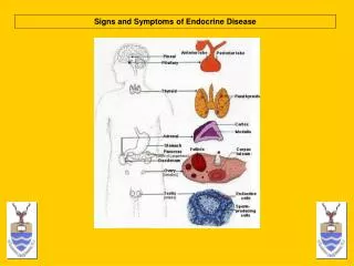
Pituitary Gland • Anterior: hormone secretion of thyroid, adrenal cortex, gonads • Posterior: water balance, salt balance
Two Major Divisions of Pituitary Anterior “Adenohypophysis” • Each has a distinct role to play in hormone regulation Posterior “Neurohypophysis”
Laboratory finding in acromegaly • Plasma glucose may be elevated • Increase serum insulin • Elevated serum phosphate • Hypercalciuria • Elevated GH
Diagnosis of acromegaly • Glucose suppression test • IGF-1 • Tumor localization MRI
Posterior pituitary hormone (ADH vasopressin) • ADH acts through tow receptors V1 @ V2 • V1 receptors mediate vascular smooth muscle contraction @stimulate prostaglandin synthesis • V2 receptors produce renal action by increase the water permeability of the luminal membrane of collecting duct epithelium • In the absence of ADH permeability of the epithelium is decrease leading to polyuria
Laboratory finding • A large urinary volume >3 l /per day • Urine osmolality less than 200 mosm/kg • Slightly elevated plasma osmolality • Low serum ADH in CDI • High or normal ADH in NDI
Diagnosis of DI • Water deprivation test (method) • Deprivation from water for 4-18 hr • Hourly measurements of urine osmolality • Continues until urine osmolality of 3 consecutive sample varies by less than 30 mosml/kg • 5 unite of AVP or 1 mg of desmopression injected @ urine and plasma osml measure 30,60,120 m later
Thyroid • Growth, development • Metabolic rate
Thyroid Function Tests • Free serum thyroxine (T4) • Free serum T3 • TSH
Scans/Ultrasound • Radioiodine uptake (RAIU) • Thyroid Scan • Ultrasound • Fine needle Aspiration
Radioiodine Uptake • Useful in differentiating non-pituitary thyrotoxic states (i.e., low TSH, high free thyroxine) • No use in hypothyroidism • A set dose of radioactive iodine (usually I123) is given and 24hrs later a radiation detector is placed over the thyroid to determine % of dose taken up by thyroid
RAIU • RAIU is increased in • Graves Disease • Hot nodules • Multi-nodular goiters • Toxic Solitary Nodule • hCG secreting tumors
RAIU • RAIU is decreased in • Amiodarone • Factitious Thyroiditis • Self limited thyroiditis-induced thyrotoxic state • Painless chronic thyroiditis • Postpartum thyroiditis • Subacute thyroiditis
Thyroid Scan • Also called scintiscan or radionuclide scan • A dose of radioiodine or Tc99m is given • Scintillation scanner produces a rough picture indicating how these isotopes localize in the thyroid • Thyroid scan is only used for nodular disease---useful for determining whether a nodule is hot or cold • Again---RAIU produces a number, scan produces a picture
Ultrasound • U/S can provide information about its size and texture • Used for determining whether a nodule is cystic or solid • Follow the size of a nodule or goiter over time.
Parathyroid gland And calcium metabolisms
CALCIUM HOMEOSTASIS DIETARY CALCIUM THE ONLY “IN” BONE DIETARY HABITS, SUPPLEMENTS ORGAN, ENDOCRINE BLOOD CALCIUM INTESTINAL ABSORPTION ORGAN PHYSIOLOGY ENDOCRINE PHYSIOLOGY KIDNEYS ORGAN PHYS. ENDOCRINE PHYS. URINE THE PRINCIPLE “OUT”
VITAMIN D SYNTHESIS SKIN LIVER KIDNEY 7-DEHYDROCHOLESTEROL VITAMIN D3 25(OH)VITAMIN D 25-HYDROXYLASE 1a-HYDROXYLASE h VITAMIND3 25(OH)VITAMIN D 1,25(OH)2 VITAMIN D (ACTIVE METABOLITE) TISSUE-SPECIFIC VITAMIN D RESPONSES
CALCIUM, PTH, AND VITAMIN D FEEDBACK LOOPS BONE RESORPTION URINARY LOSS 1,25(OH)2 D PRODUCTION SUPPRESS PTH RISING BLOOD Ca NORMALBLOOD Ca FALLING BLOOD Ca BONE RESORPTION URINARY LOSS 1,25(OH)2 D PRODUCTION STIMULATE PTH
Synthesis of Adrenocortical Hormones • Zona glomerulosa: aldosterone • Zona fasciculata: glucocorticoids (cortisol) • Zona reticularis: androgens (DHEA and androstenedione)
Regulation of Adrenocortical Hormones • Hypothalamus • CRH-containing neurons are stimulated • CRH delivered to anterior pituitary • CRH binds to receptors on corticotrophs, causing synthesis and secretion of ACTH
Cushing’s Syndrome • Diagnosis of Cushing’s syndrome • History • Weight gain, fatigue • Infertility, impotence, changes in menstruation • Diabetes, polyuria, polydypsia • Depression, headache • Signs of underlying tumor (weight loss, appetite) • Physical exam • Obesity, fat distribution • Proximal muscle weakness/wasting • Palpation of abdominal mass
Cushing’s Syndrome • Diagnosis of Cushing’s syndrome • Labs • 24 hour urinary cortisol • 2-3 consecutive days • Verify with creatinine values • Spot AM/PM serum cortisol • Circadian variation • AM ACTH surge causes increased cortisol • PM should see at least 50% drop in cortisol level • Low-dose dexamethasone suppression test
Cushing’s Syndrome • Diagnose cause of Cushing’s syndrome • History (steroid use?) • Serum ACTH • Elevated : Cushing’s disease, ectopic ACTH • Suppressed: primary adrenal source • Correlate with cortisol levels • High-dose dexamethasone suppression test • Metyrapone test
Cushing’s Syndrome • Dexmethasone suppression test • Synthetic glucocorticoid (30x more potent as inhibitor) • Low dose • 0.5mg po q6 hours x48 hours • Measure cortisol, 17-hydroxycorticosteroid, creatinine • Fall in all steroid levels in pseudo-Cushing and normals • Differentiates presence/absence of Cushing’s syndrome • Alternative dosing • 1mg po at midnight and measure 8am cortisol • Much less sensitive
Cushing’s Syndrome • Dexmethasone suppression test • High Dose • 2mg po q6 hours x48 hours • Measure cortisol and urinary free cortisol • Ectopic ACTH and adrenal tumors- no suppression • Cushing’s disease- suppress to <50% of baseline • Usually only used if ACTH/Cortisol assays unavailable or equivocal
Cushing’s Syndrome • Metyrapone test • Inhibits 11-B-hydroxylase • Blocks conversion of 11-deoxycortisol to cortisol • Plasma cortisol levels fall and ACTH increases • Marked increase in 17-hydroxycorticosteroid levels and 11-deoxycortisol levels • Cushing’s Disease- normal or supernormal increase in levels • Ectopic ACTH or adrenal sources- no response • Risks adrenal insufficiency
Cushing’s Syndrome • Petrosal vein sampling • Measure petrosal venous sinus ACTH level and correlate to plasma levels • Invasive with morbidity • Usually not used • Adrenal venous sampling • Measure cortisol and aldosterone • Not used anymore
Cushing’s Syndrome • Radiographic Localization • CT of sella turcica • Unenhanced and gadolinium enhanced MRI • Radionuclide imaging for somatostatin receptors • >60% sensitive • 1st study if diagnosed with Cushing’s syndrome • CT of chest/abdomen with 3mm cuts through adrenal • Adrenal hyperplasia • Thickening and elongation of adrenal rami bilaterally • Multinodularity of cortex bilaterally
Cushing’s Syndrome • Radiographic Localization • CT of adrenal glands • Adenomas- usually >2cm but <5cm • Low attenuation (lipid content) • Atrophy of opposite gland • Carcinoma- indistinguishable from adenomas • >5cm • Necrosis, calcifications, irregularity, invasion • MRI of adrenal- usually not needed • Signal intensity much higher than in spleen = carcinoma • Adjacent organ and/or vascular involvement
Cushing’s Syndrome • Radiographic Localization • CT of adrenal glands • Adenomas- usually >2cm but <5cm • Low attenuation (lipid content) • Atrophy of opposite gland • Carcinoma- indistinguishable from adenomas • >5cm • Necrosis, calcifications, irregularity, invasion • MRI of adrenal- usually not needed • Signal intensity much higher than in spleen = carcinoma • Adjacent organ and/or vascular involvement
DIAGNOSIS • Basal Cortisol Level • Avoid random level: low sensitivity • Check morning cortisol • Greater than 18 µg/dL indicates an intact axis • Less than 3 µg/dL strongly suggests insufficiency • Intermediate values: perform cosyntropin stimulation test
DIAGNOSIS • Low-Dose Cosyntropin Test • Cosyntropin doses as low as 0.5 to 1 ug will give a maximal response within 15 to 30 mins • Possibly superior to high-dose test for diagnosing secondary insufficiency because ACTH level closer to physiologic level • Normal response: peak plasma cortisol level > 18 µg/dL • Like high-dose test, low sensitivity when ruling out mild or recent secondary insufficiency; confirm with more sensitive tests (insulin tolerance, metyrapone)
DIAGNOSIS • Insulin Tolerance Test • Hypoglycemia induced by the IV injection of reg insulin stimulates the entire HPA axis. • Plasma glucose and cortisol are measured after injection of insulin. • Normal response: cortisol increases to at least 18ug per dL • Expensive and contraindicated in patients with coronary heart disease or seizures
DIAGNOSIS • Metyrapone Test • Metyrapone inhibits conversion of 11-deoxycortisol to cortisol • Give at midnight and measure the concentration of 11-deoxycortisol and cortisol at 8am • In normal subjects, the plasma 11-deoxycortisol concentration increases to at least 7ug per dL. In patients with adrenal insufficiency, the increase is smaller and is related to the severity of the corticotropin deficiency
Hyperaldosteronism • Causes • Adenoma (most common) • F>M • 4th-5th decades of life • Bilateral adrenal hyperplasia • Adrenocortical carcinoma (rare) • Glucocorticoid remedial aldosteronism • Aldosterone producing adenoma • Responsive to renin
Hyperaldosteronism • Diagnosis • History, HTN, symptoms • Laboratory • Serum K+ (<3.0) • Serum aldosterone • Salt load patients (suppresses aldosterone) • Level >14 micrograms/d diagnostic of primary hyperaldosteronism • Serum renin • If >1.0 then unlikely primary hyperaldosteronism