Download
1 / 33
380 likes | 1.17k Views
Partograph. A partograph is a graphical record of the observations made of a women in labour For progress of labour and salient conditions of the mother and fetus It was developed and extensively tested by the world health organization WHO. History Of Partogram.

E N D
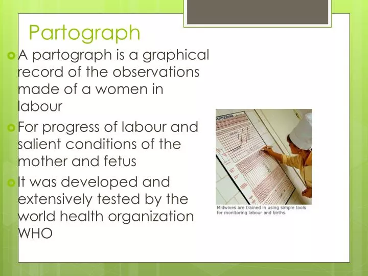
Partograph • A partograph is a graphical record of the observations made of a women in labour • For progress of labour and salient conditions of the mother and fetus • It was developed and extensively tested by the world health organization WHO
History Of Partogram • Friedman's partogram devised in 1954 was based on observations of cervical dilatation and fetal station against time elapsed in hours from onset of labour. The time onset of labour was based on the patient's subjective perception of her contractility. Plotting cervical dilatation against time yielded the typical sigmoid or 'S' shaped curve and station against time gave rise to the hyperbolic curve. Limits of normal were defined
Overview • The partograph can be used by health workers with adequate training in midwifery who are able to : - observe and conduct normal labour and delivery. - Perform vaginal examination in labour and assess cervical diltation accurately - plot cervical diltation accurately on a graph against time • There is no place for partograph in deliveries at home conducted by attendants other than those trained in midwifery • Whether used in health centers or in hospitals , the partograph must be accompanied by a program of training in its use and by appropriate supervision and follow up
Objectives • early detection of abnormal progress of a labour • prevention of prolonged labour • recognize cephalopelvic disproportion long before obstructed labour • assist in early decision on transfer , augmentation , or termination of labour • increase the quality and regularity of all observations of mother and fetus • early recognition of maternal or fetal problems • the partograph can be highly effective in reducing complications from prolonged labor for the mother (postpartum hemorrhage, sepsis, uterine rupture and its sequelae) and for the newborn (death, anoxia, infections, etc.).
Partograph function • The partograph is designed for use in all maternity settings , but has a different level of function at different levels of health care • in health center, the partograph,s critical function is to give early warning if labour is likely to be prolonged and to indicate that the woman should be transferred to hospital (ALERT LINE FUNCTION ) • in hospital settings, moving to the right of alert line serves as a warning for extra vigilance , but the action line is the critical point at which specific management decisions must be made • other observations on the progress of labour are also recorded on the partograph and are essential features in management of labour
Components of the partograph • Part 1 : fetal condition ( at top ) • Pqrt 11 : progress of labour ( at middle ) • Part 111 : maternal condition ( at bottom ) • Outcome : ………………
Part 1 : Fetal condition • this part of the graph is used to monitor and assess fetal condition • 1 - Fetal heart rate • 2 - membranes and liquor • 3 - moulding the fetal skull bones • Caput
Fetal heart rate Basal fetal heart rate? • < 160 beats/mi =tachycardia • > 120 beats/min = bradycardia • >100beats/min = severe bradycardia Decelerations? yes/no Relation to contractions? • Early • Variable • Late – -----Auscultation - return to baseline > 30 sec contraction ----- Electronic monitoring peak and trough (nadir) > 30 sec
membranes and liquor • intact membranes ……………………………………….I • ruptured membranes + clear liquor …………………….C • ruptured membranes + meconium- stained liquor ……..M • ruptured membranes + blood – stained liquor …………B • ruptured membranes + absent liquor…………………....A
moulding the fetal skull bones • Molding is an important indication of how adequately the pelvis can accommodate the fetal head • increasing molding with the head high in the pelvis is an ominous sign of cephalopelvic disproportion • separated bones . sutures felt easily ……………….….O • bones just touching each other ………………………..+ • overlapping bones ( reducible 0 ……………………...++ • severely overlapping bones ( non – reducible ) ……..+++
part11 – progress of labour . Cervical diltation • Descent of the fetal head • Fetal position • Uterine contractions • this section of the paragraph has as its central feature a graph of cervical diltation against time • it is divided into a latent phase and an active phase
latent phase : • it starts from onset of labour until the cervix reaches 3 cm diltation • once 3 cm diltation is reached , labour enters the active phase • lasts 8 hours or less • each lasting < 20 sceonds • at least 2/10 min contractions
Active phase : • Contractions at least 3 / 10 min • each lasting < 40 sceonds • The cervix should dilate at a rate of 1 cm / hour or faster
Alert line ( health facility line ) • The alert line drawn from 3 cm diltation represents the rate of diltation of 1 cm / hour • Moving to the right or the alert line means referral to hospital for extra vigilance
Action line ( hospital line ) • The action line is drawn 4 hour to the right of the alert line and parallel to it • This is the critical line at which specific management decisions must be made at the hospital
Cervical diltation • It is the most important information and the surest way to assess progress of labour , even though other findings discovered on vaginal examination are also important • when progress of labour is normal and satisfactory , plotting of cervical dilatation remains on the alert line or to left of it • if a woman arrives in the active phase of labour , recording of cervical dilatation starts on the alert line • when the active phase of labor begins , all recordings are transferred and start by plotting cervical dilatation on the alert line
Descent of the fetal head • It should be assessed by abdominal examination immediately before doing a vaginal examination, using the rule of fifth to assess engagement • The rule of fifth means the palpable fifth of the fetal head are felt by abdominal examination to be above the level of symphysis pubis • When 2/5 or less of fetal head is felt above the level of symphysis pubis , this means that the head is engage , and by vaginal examination , the lowest part of vertex has passed or is at the level of ischial spines
Assessing descent of the fetal head by vaginal examination; 0 station is at the level of the ischial spine (Sp).
Fetal position Occiput transverse positions Occiput anterior positions
Uterine contractions • Observations of the contractions are made every hour in the latent phase and every half-hour in the active phase • frequency how often are they felt ? • Assessed by number of contractions in a 10 minutes period • duration how long do they last ? Measured in seconds from the time the contraction is first felt abdominally , to the time the contraction phases off • Each square represents one contraction
Palpate number of contraction in ten minutes and duration of each contraction in seconds • Less than 20 seconds: • Between 20 and 40 seconds: • More than 40 seconds:
Part111: maternal condition Name / DOB /Gestation Medical / Obstetrical issues Assess maternal condition regularly by monitoring : • drugs , IV fluids , and oxytocin , if labour is augmented • pulse , blood pressure • Temperature • Urine volume , analysis for protein and acetone
- latant phase is less than 8 hours- progress in active phase remains on or left of the alert line • Do not augment with oxytocin if latent and active phases go normally • Do not intervene unless complications develop • Artificial rupture of membranes ( ARM ) • No ARM in latent phase • ARM at any time in active phase
Between alert and action lines • In health center , the women must be transferred to a hospital with facilities for cesarean section , unless the cervix is almost fully dilated • Observe labor progress for short period before transfer • Continue routine observations • ARM may be performed if membranes are still intact
At or beyond action line • Conduct full medical assessement • Consider intravenous infusion / bladder catheterization / analgesia • Options - Deliver by cesarean section if there is fetal distress or obstructed labour - Augment with oxytocin by intravenous infusion if there are no contraindications
Moving to the right of alert line • This means warning • Transfer the woman from health center to hospital • reaching the action line • This means possible danger • Decision needed on future management (usually by obesteritian or resident )
Prolonged latent phase • If a woman is admitted in labor in the latent phase ( less than 3 cm diltation ) and remains in the latent phase for next 8 hours • Progress is abnormal and she must br transferred to a hospital for a decision about further action • This is why there is a heavy line drawn on the partograph at the end of 8 hours of the latent phase
Polonged Active phase • In the active phase of labor , plotting of cervical diltation will normally remain on or to the left of the alert line • But some cases will move to the right of the alert line and this warns that labor may be prolonged • This will happen if the rate of cervical diltation in the active phase of labor is not 1 cm / hour or faster • A woman whose cervical diltation moves to the right of the alert line must be transferred and manged in a hospital with adequate facilities for obstetric intervention unless delivery is near • at the action line , the woman must be carefully reassessed for why labor is not progressing and a decision made on further management
Secondary arrest of cervical diltation • Abnormal progress of labor may occur in cases with normal progress of cervical diltation then followed by secondary arrest of diltation
Secondary arrest of head descant • Abnormal progress of labor may occur with normal progress of descent of the fetal head then followed by secondary arrest of descent of fetal head