Download
1 / 47
490 likes | 820 Views
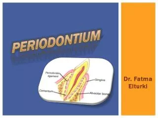
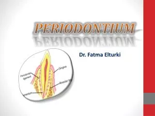
Tour of the Periodontium. Jafar Naghshbandi D.D.S;M.S Diplomate of the American Board of Periodontology. Special Thanks Whoever taught me a word made me his servant. Dr. Jim Simon. Dr. Raul Caffesse. INTRODUCTION. Periodontium supporting structures. Gingiva. GINGIVA.

E N D
Tour of the Periodontium JafarNaghshbandi D.D.S;M.S Diplomate of the American Board of Periodontology
Special ThanksWhoever taught me a word made me his servant. Dr. Jim Simon Dr. Raul Caffesse
GINGIVA • Part of oral mucosa that covers the alveolar processes of the jaws and surrounds the necks of the teeth. • Marginal gingiva • A. Gingival Sulcus • B. Interdental Gingiva
Marginal Gingiva • Terminal edge of the Gingiva surrounding the teeth in collar like fashion. • 1.5-2mm wide • Channel Way Gingival Sulcus • V-Shaped shallow Crevice • 1.8mm • Presence of crevicular fluid
Interdental gingiva • Occupy gingival embrasure • Site of initial lesion on gingivitis • Anterior region-Pyramidal form and flattened over Molar region • Scalloped Form • Presence of Interdental COL
Attached Gingiva • Functional Mucosa • Firm, resilient and tightly bound to alveolar Bone • Pale, pink and Stippled appearance
periodontal ligament • “The is the connective tissue that surrounds the root & connects it to the bone. It is continuous with the connective tissue of the gingiva & communicates with the marrow spaces through vascular channels in the bone.” • Carranza & Bernard
periodontal ligament • Periodontal Ligament Surrounds the root of the tooth • Composed of fibers, or ligaments, that support and suspend the tooth in the socket • Fibers are arranged in bundles • Forms a ‘shock-absorber’ for the tooth in the socket
periodontal ligament SHAPE • It is thinnest around the middle third of the root, with an hour glass appearance. • The ligament appears as a radiolucent area of 0.4- 1.5mm between the radiopaque lamina dura of the alveolar bone and cementum.
periodontal ligament AVERAGE WIDTH : • Depending on age • 11-16 yrs - 0.21mm • 32-52 yrs - 0.18mm • 51-67 yrs - 0.15mm • According to functional state of the tissues • Time of eruption - 0.1- 0.5mm • At function - 0.2-0.35mm • Hypo function - 0.1-0.15mm 9/71
Cementum • Definition: It is a mineralized dental tissue covering the anatomic roots of human teeth • It was first demonstrated microscopically in 1835by pupils of Purkinje • The word cementum comes from the latin word “cement” which means quarried stone
Cementum PHYSICAL CHARACTERISTICS • Calcified structure whose calcification and hardness is less than dentin • More permeable than dentin • Light yellow in color • Softer and lighter than dentin • Lacks luster and is dark, and is therefore differentiated from enamel
Cementum • Less readily resorbed than bone EXTENSION: • Begins at the cervical portion of the tooth at the CEJ and continues to the apex T THICKNESS: • At CEJ : 10 micrgons ( thinnest ) • At Apical Region : 200-300 microns ( thickest )
Cementum EXTENSION: • Begins at the cervical portion of the tooth at the CEJ and continues to the apex THICKNESS: • At CEJ : 10 microns ( thinnest ) • At Apical Region : 200-300 microns ( thickest )
Cementum Classification of cementum Based upon A. Location B. Presence or absence of cells C. Origin of collagenous fibers of the matrix D. Acc to Schoreder
Cementum B) Presence or absence of cells I. Cellular II. Acellular
Cemento-Enamel Junction • Cementum overlaps enamel – 60% • Cementum just meets enamel – 30% • Small gap between cementum and enamel – 10%
Injuries to Cementum VERTICAL FRACTURE • Poor prognosis and usually it cannot be repaired by cementum easily • Treatment :extraction or stabilization by intracoronal splinting HORIZONTAL FRACTURE • prognosis depends on the age and location of fracture. • Apical or middle third: it can be repaired by the cementum and prognosis for the vitality of the pulp of the tooth for survival is fair • The coronal third: prognosis for vitality of tooth is poor
Cementum Repair ANATOMIC REPAIR : • Generally occurs when the degree of destruction is low • The root outline is re- established as it was before cemental resorption FUNCTIONAL REPAIR : • Occurs in cases of large cemental resorption or destruction • To maintain the width of periodontal ligament, the adjacent alveolar bone grows and takes the shape of defect following the root surface. This is done to improve the function of tooth
Alveolar Bone • Also called as alveolar process • Definition: alveolar process is that part of maxilla and mandible that forms and supports the sockets of teeth • It is composed mainly of two parts: • Alveolar bone proper • Supporting bone • When the teeth are lost the alveolar process disappears
structure of alveolar bone A) Alveolar bone proper • lamellated bone • bundle bone B) Supporting alveolar bone • Cortical plate • Spongy bone
ALVEOLAR BONE PROPER • It surrounds the root of the tooth and gives attachment to the periodontal ligament fibers. It consists of: • Lamellated bone • Bundle bone
lamina dura The alveolar process contains a region of compact bone adjacent to the periodontal ligament called Lamina dura It is the bone lining the alveolus which is attached to the cementum of the roots by the periodontal ligament In clinical radiographs, it normally appears as a dense white line
Alveolar Socket • Also called Dental alveolus • Sockets in the jaws in which the roots of teeth are held in the alveolar process with the periodontal ligament • Alveolar socket of the second premolar tooth in a bovine maxillary bone.
Septa • “Septa” – in Latin, it means “fence” or “wall” • Interdental Septa • Are plates of bone that separate each individual sockets from one another • Interradicular Septa • Are thin plates of bone that separate the roots of multi-rooted teeth