Download
1 / 29
450 likes | 1.37k Views
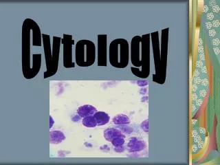
Vaginal Cytology Evaluation. Clinical Pathology. Vaginal Cytology Indications. Examine exfoliated cells from the vagina Vaginal epithelium is ovarian hormonal influenced- stages estrus for optimum breeding time. Knowledge of the onset of vaginal discharge Character of the discharge

E N D
Vaginal Cytology Evaluation Clinical Pathology
Vaginal Cytology Indications • Examine exfoliated cells from the vagina • Vaginal epithelium is ovarian hormonal influenced- stages estrus for optimum breeding time. • Knowledge of the onset of vaginal discharge • Character of the discharge • Degree of vulvar swelling • Attitude of female towards male dog • Also useful in detecting inflammation and neoplasia of the genital tract.
Vaginal Cytology Technique • May or may not want to use a speculum (laryngoscope may work as well). • Carefully part labia • Direct swab craniodorsally • Swab the vaginal wall • Avoid the vestibule and clitorial fossa, superficial cells may alter results • Roll on glass slide, let dry and stain (Diff-Quik)
http://www.vetmed.lsu.edu/eilts/Multimediafiles/Sound/Video/Canine%20Cytology.mpghttp://www.vetmed.lsu.edu/eilts/Multimediafiles/Sound/Video/Canine%20Cytology.mpg
Vaginal Epithelial Cell Types • Basal Cells (may not be seen) • Parabasal Cells • Intermediate Cells • Superficial Cells
Parabasal Cells • Small round cells with round nuclei and small amount of cytoplasm • Uniform in size and shape
Intermediate Cells • May be small or large • Round nuclei, nucleus similar in size as parabasal cells • Entire cell approximately twice the size of parabasal cells • Cytoplasm becomes angular, irregular and folded as cell enlarges
Superficial Cells • Largest epithelial cell • As they age and degenerate, the nuclei becomes small, pyknotic and fades. • Cytoplasm may contain vacuoles with age
Superficial Cells Continued • Cornification is the degeneration process • Superficial cells are commonly called cornified cells • Once nucleus is lost become Anuclear cells
Staging the Canine Estrus Cycle • Stages of Estrus • Proestrus • Estrus • Diestrus • Anestrus
Proestrus • Bitch has a swollen vulva, reddish vulvar discharge • Will not accept the male during this time • Duration is 9 days (with possibility of 2-15) • Parabasal cells and intermediate cells gradually decrease in number as the cycle nears estrus • Superficial cells appear by 2-3 day and increase in number over time • Erythrocytes are numerous and gradually decline • Neutrophils are also present and decline
Early Proestrus • Parabasal and intermediate cells predominate • As proestrus progresses, parabasal cells disappear as superficial cells increase.
Late Proestrus • White Blood cells decrease in number • Large intermediate and superficial cells predominate • No parabasal cells or small intermediate cells • Red blood cells or present or absent • Bacteria is often present
Estrus • Lasts an average of 9 days • Female accepts male • Vulvar discharge is less bloody • Vulva is softer • Sometimes bloody discharge may continue through estrus
Estrus • Superficial cells predominate (90%) • May become cornified • NO WBC’S EXCEPT AT LAST 1-2 DAYS OF ESTRUS!!!!!!!!!!!!!!!!!!!!!!!! • Variable RBC’s • Large numbers of bacteria
Estrus-Hormonal Events • Serum progesterone increases above anestrus range • Progesterone rise begins when LH peaks • Ovulation occurs 2 days after LH peak • Eggs take an additional 2-3 days to mature • Fertile period is 4-7 days after LH peak • Now do blood tests to check for LH peak
Diestrus • Abrupt decrease in superficial cells • Increase in parabasal cells and intermediate cells • Many WBC’s, then decrease in late diestrus • Variable RBC’s • Occurs average 8 days after LH peak • Lasts about 2 months • Progesterone peaks 15-30 days post estrus, then declines
Anestrus • The transition period between cycles • 4-12 months • Parabasa and intermediate cells predominate • Few WBC’s and bacteria
Feline Vaginal Cytology • More difficult to collect • Ovulation may be induced due to stimulation of cervix during collection • Similar to a dog, but develop at a faster rate. Average estrus is 8 days, interval between estrus period is 9 days if ovulation does not occur. • Usually no vaginal or scant vaginal discharge may be seen • Usually no RBC’s are present during proestrus
Management of Breeding • Do comparison cytology during late proestrus and estrus • Breed every other day beginning on day 9 • Use other hormonal test kits to help stage cycle • Progesterone ELISA Assay • LH Assay