Download
1 / 27
360 likes | 928 Views
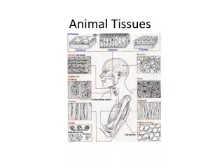
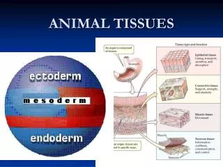
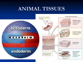
Lab 1 ANIMAL TISSUES. Animals are multicellular heterotrophs whose cells lack cell walls. Most animals exhibit a hierarchical level of organization: Cells are organized into tissues Tissues combine to form organs Organs comprise organ systems. Levels of Organization.

E N D
Animals are multicellularheterotrophs whose cells lack cell walls. Most animals exhibit a hierarchical level of organization: • Cells are organized into tissues • Tissues combine to form organs • Organs comprise organ systems Levels of Organization
Group of similar cells that perform a specialized function. Examples include: • Bone tissue • Blood tissue • Muscle tissue What is a tissue?
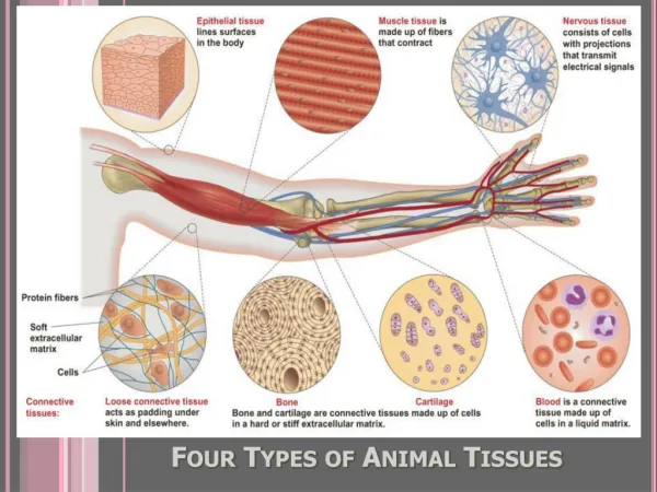
Epithelial • Connective • Muscle • Nervous 4 basic types of animal tissue:
Characteristics: • Cells fit closely together forming continuous sheets Epithelial Tissue • Apical (free) surface covers body surface or lines interior of organs • Basal surface adheres to the basement membrane
Supported by connective tissue • Avascular, but innervated • Have remarkable powers of regeneration Variety of functions depending on type (protection, absorption, filtration, excretion, secretion) Epithelial Tissue
Classification based on # of cell layers and shape of cells on apical surface. • # of Cell Layers: • Simple – one layer of cells • Stratified – two or more layers • Pseudostratified – simple, but appears stratified Epithelial Tissue
Cell shape on apical surface: • Squamous – flattened & scale-like • Cuboidal – box-like • Columnar – tall & column-like Epithelial Tissue
Characteristics: • Most are well vascularized • Consists of widely-spaced cells and fibers embedded in a non-living extracellular matrix Variety of functions depending on type (support, binding other tissues, transport, defense, storage) Connective Tissue
Characteristics: • Well vascularized • Packed with actin & myosin filaments Function to contract producing most types of body movements Muscle Tissue
Characteristics: • Composed of two types of cells: • Neurons – specialized to generate and transmit impulses; amitotic • Neuroglia (glial cells) – protect, support & insulate neurons • Main component of the nervous system (brain, spinal cord & nerves) Nervous Tissue
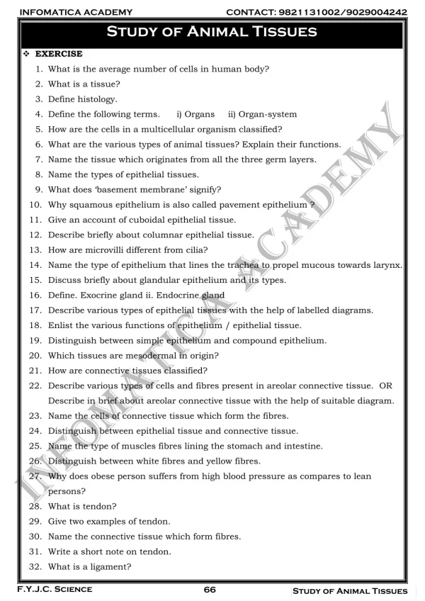
This week’s lab is devoted to histology(the study of tissues).
Simple squamous epithelium • Location – alveoli of lungs, lining of heart & blood vessels • Function – allows diffusion of materials Exercise A: Epithelial Tissues surface view lateral view
Simple cuboidal epithelium • Location – kidney tubules & ducts; ovary surface • Function – secretion & absorption Exercise A: Epithelial Tissues simple cuboidal epithelium basement membrane cross section longitudinal section
Simple columnar epithelium • Location – lines digestive tract from stomach to the rectum • Function – absorption & secretion Exercise A: Epithelial Tissues
Pseudostratified columnar epithelium • Location – lining of trachea & upper respiratory tract • Function – secretion & propulsion of mucus Exercise A: Epithelial Tissues cilia ciliated cell goblet cell basal cell basement membrane connective tissue
Stratified squamous epithelium • Location • keratinized type: epidermis of skin • non-keratinized type: linings of esophagus, mouth & vagina • Function – protection Exercise A: Epithelial Tissues stratified squamous epithelium connective tissue Non-keratinized Keratinized
Loose (areolar) connective tissue • Location – widely distributed under epithelia • Function – cushions organs Exercise B: Connective Tissues Gel-like matrix Collagen fiber Fibroblast nucleus Elastic fiber
Adipose • Location – under skin; around kidneys & eyeballs; in breasts • Function – supports & protects organs; insulates against heat loss; provides reserve fuel Exercise B: Connective Tissues
Dense (fibrous) connective tissue • Location – tendons & ligaments • Function – attaches muscle to bone (tendons) & bone to bone (ligaments) Exercise B: Connective Tissues
Hyaline cartilage • Location – covers ends of long bones; nose, trachea & larynx • Function – support & reinforcement Exercise B: Connective Tissues Chondrocytes sitting in lacunae (cavities) Matrix packed with collagen fibers
Bone • Location – bones • Function – support & protection; calcium storage; provides levers for muscles to act on; site of blood cell production Exercise B: Connective Tissues Canaliculi Central canal Osteocyte sitting a in lacuna (cavity) Osteon
Blood • Location – contained within blood vessels • Function – transport of gases (O2 & CO2), nutrients & metabolic wastes Exercise B: Connective Tissues Red blood cells Platelet • White blood cells: • neutrophil • monocyte • lymphocyte Plasma (liquid matrix)
Skeletal muscle • Long, cylindrical, multinucleate cells with obvious striations • Location – attached to bones or occasionally to skin • Function – voluntary movement Exercise C: Muscle Tissues Nuclei Striations
Cardiac muscle • Branched, uninucleate cells with striations • Location – walls of the heart • Function – contract involuntarily to propel blood Exercise C: Muscle Tissues Branched cell Intercalated disc Striations
Smooth muscle • Tapered, uninucleate, non-striated cells • Location – walls of hollow organs • Function – contract involuntarily to propel materials along internal passageways Exercise C: Muscle Tissues Nuclei Circular layer Longitudinal layer Individual muscle cell
Neurons • cell body – contains nucleus • cytoplasmic processes: • dendrites – transmit impulses to cell body • axon – transmits impulses from cell body Exercise D: Nervous Tissue Neuronal processes (axons & dendrites) Neuron nucleus Neuroglia Neuron cell body