Download
1 / 57
570 likes | 943 Views
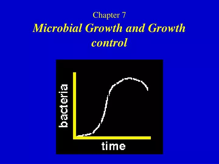
Chapter 7 Microbial Growth and Growth control. Chapter outline. 7.1 Overview of Cell Growth 7.2 Population Growth 7.3 Measurement of Growth 7.4 Continuous Culture: The Chemostat 7.5 Effect of Environment on Growth 7.6 Growth Control 7.7 Viral Control 7.8 Fungal Control. Concepts.

E N D
Chapter outline 7.1 Overview of Cell Growth 7.2 Population Growth 7.3 Measurement of Growth 7.4 Continuous Culture: The Chemostat 7.5 Effect of Environment on Growth 7.6 Growth Control 7.7 Viral Control 7.8 Fungal Control
Concepts • Microbial growth is defined as an increase in cellular constituents and may result in an increase in a microorganism’s size, population number, or both. • A wide variety of techniques can be used to study microbial growth by following changes in the total cell number, the population of viable microorganisms, or the cell mass. • Solid objects can be sterilized by physical agents such as heat and radiation; Liquids and gases are sterilized by heat, radiation, and filtration through the proper filter. • A knowledge of methods used for microbial control is essential for personal and public safety.
7.1 Overview of microbial growth • The bacterial cell is a synthetic machine that is able to duplicate itself. The processes involve as many as 2000 chemical reactions of a wide variety of types: • Energy transformations. • Biosynthesis of small molecules--the building, blocks of macromolecules-as well as the various cofactors and coenzymes needed for enzymatic reactions.
Growth definition Growth may be generally defined as a steady increase in all of the chemical components of an organism. Growth usually results in an increase in the size of a cell and frequently results in cell division.
Cell life cycle in Eukaryotic cells G1 Primary growth phase of the cell during which cell enlargement occurs, a gap phase separating cell growth from replication of the genome SPhase in which replication of the genome occurs G2Phase in which the cell prepares for separation of the replicated genomes, this phase includes synthesis of microtubules and condensation of DNA to form coherent chromosomes, a gap phase separating chromosome replication from miosis. M Phase called miosis during which the microtubular apparatus is associated and subsequently used to pull apart the sister chromosomes.
Eukaryotic cell: Prokaryotic cell: G1 S G2 M G1R D
Binary fision • Most bacterial cells reproduce asexually by binary fision, a process in which a cell divides to produce two nearly equal-sized progeny cells. • Three processes: • Increase in cell size (cell elongation) • DNA replication • Cell division
7.2 Population Growth • Growth is defined as an increase in the number of microbial cells in a population. • Growth rate is the change in cell number or cell mass per unit time. • The interval for the formation of two cells from one is called a generation. • The time required for this to occur is called the generation time.
Exponential Growth A growth experiment beginning with a single cell having a doubling time of 30 min is presented. This pattern of population increase, where the number of cells doubles during each unit time period, is referred to as exponential growth.
Calculating Generation Times • N = N02n N = final cell number. N0 = initial cell number, and n = number of generations. • n= ( log N0 − log N ) /log 2 = ( log N0 − log N ) / 0.301= 3.3 (log N− log N0) . • The generation time g of the cell population is calculated as t / n, where t is simply the hours or minutes of exponential growth.
Growth Cycle of Populations A typical growth curve for a population of cells can be divided into several distinct phases called the lag phase, exponential phase, stationary phase, and death phase.
Growth curve of bacteria • Lag Phase • Exponential Phase • Stationary Phase • Death Phase
Lag Phase When a microbial population is inoculated into a fresh medium, growth usually does not begin immediately but only after a period of time called the lag phase, which may be brief or extended depending on the history of the culture and growth conditions. This happens because for growth to occur in a particular culture medium the cells must have a complete complement of enzymes for synthesis of the essential metabolites not present in that medium.
Exponential Phase It is a consequence of the fact that each cell divides to form two cells, each of which also divides to form two more cells, and so on. Most unicellular microorganisms grow exponentially, but rates of exponential growth vary greatly. In general, prokaryotes grow faster than eukaryotic microorganisms
Stationary Phase If a single bacterium continued to grow exponentially for 48 hr, produce a population that weighed about 4000 times the weight of Earth! This is particularly impressive because a single bacterial cell weighs only about one-trillionth (10− l2) of a gram. An essential nutrient of the culture medium is used up or some waste product of the organism builds up in the medium to an inhibitory level and exponential growth ceases,or both.
Death Phase If incubation continues after a population reaches the stationary phase, the cells may remain alive and continue to metabolize, but they may also die. If the latter occurs, the population is said to be in the death phase.
7.3 Measurement of Growth Population growth is measured by following changes in the number of cells or weight of cell mass.
Total Cell Count The number of cells in a population can be measured by counting a sample under the microscope, either on samples dried on slides or on samples in liquid. With liquid samples, special counting chambers must be used. Direct microsopic counting procedure using the Petroff-Hausser counting chamber.
Viable Counting method The usual practice, which is the most valid statistically, is to count colonies only on plates that have between 30 and 300 colonies. To make a 10-fold (10−1) dilution, one can mix 0.5 ml of sample with 4.5 ml of diluent, or 1.0 ml sample with 9.0 ml diluent.
The usual way to perform a viable count is to determine the number of cells in the sample capable of forming colonies on a suitable agar medium. There are two ways of performing a plate count: the spread plate method and the pour plate method.In either case the sample must usually be diluted before plating.
Sources of Error in Plate Counting • The number of colonies obtained in a viable count depends not only on the inoculum size but also on the suitability of the culture medium and the incubation conditions used; It also depends on the length of incubation. • The length of incubation. • Some tiny colonies may be missed during the counting. • The incubation conditions (medium, temperature, time). • Key dilutions must be prepared.
Turbidimetric Measurements of Cell Number A cell suspension looks cloudy (turbid) to the eye because cells scatter light passing through the suspension. The more cells present, the more light scattered and hence the more turbid the suspension.
7.4 Continuous Culture: The Chemostat A continuous culture is essentially a flow system of constant volume to which medium is added continuously and from which continuous removal of any overflow can occur. Once such a system is in equilibrium, cell number and nutrient status remain constant, and the system is said to be in steady state.
The Chemostat The most common type of continuous culture device used is a chemostat, which permits control of both the population density and the growth rate of the culture. Both parameters can be set by the experimenter.
Continuous culture of microorganisms Chemostat used for continuous cultures. Rate of growth can be controlled either by controlling the rate at which new medium enters the growth chamber or by limiting a required growth factor in the medium.
Relationship among nutrient concentration, growth rate(solid line),and growth yield(dashed line)in a batch culture(closed system). At low nutrient concentrations both growth rate and growth yield are affected.
Steady-state relationships in the chemostat. The dilution rate is determined from the flow rate and the volume of the culture vessel.Thus,with a vessel of 1000ml and a flow rate through the vessel of 500ml/hr, the dilution rate would be 0.5hr-1. Note that at high dilution rates, growth cannot balance dilution, and the population washes out. Note also that although the population density remains constant during steady state, the growth rate (doubling time)can vary over a wide range. Thus, the experimenter can obtain populations with widely varying growth rates without affecting population density.
Experimental Uses of the Chemostat • Even over rather wide ranges, any desired growth rate can be obtained in the chemostat by simply varying the dilution rate. • A practical advantage to the chemostat is that a population may be maintained in the exponential growth phase for long periods, for days and even weeks. The experimenter using the chemostat can have such cells available at any time.
7.5 Effect of Environment on Growth • The activities of microorganisms are greatly affected by the chemical and physical conditions of their environments. Understanding environmental in fluences helps us to explain the distribution of microorganisms in nature and makes it possible for us to devise methods for controlling microbial activities and destroying undesirable organisms. • Four main factors : Temperature, pH, water availability, and oxygen.
Effect of temperature on bacterial growth rate Bacteria grow over a range of temperatures; they do not reproduce below the minimum growth temperattire nor above the maximum growth temperature. Within the temperature growth range there is an optimum growth temperature at which bacterial reproduction is fastest.
Enzymes exhibit a Q10 so that within a suitable temperature range the rate of enzyme activity doubles for every 10' C rise in temperature.
Microorganisms are classified as psychrophiles, mesophiles.thermophiles, and extremethemophiles based on their optimal growth temperature.
Effect of oxygen concentration on the growth of various bacteria in tubes of solid medium. (b) Facultative anaerobes growth is best near the surface, where oxygen is available, but occurs throughout the tube. (a) Obligate aerobes-growth occurs only in the short distance to which the oxygen diffuses into the medium. (e) Microaerophiles, aerobic organisms that do not tolerate atmospheric concentrations of oxygen-growth occurs only in a narrow band of optimal oxygen concentration (c) Obligate anaerobes-oxygen is toxic, and there is no growth near the surface. (d) Aerotolerant anaerobes-growth occurs evenly throughout the tube but is not better at the surface because the organisms do not use oxygen.
Bacteria Neutral condition Fungi Acidic condition Actinomycetes Alkaline condition
Water activity The water activity of a solution is 1/100 the relative humidity of the solution (when expressed as a percent), or it is equivalent to the ratio of the solution's vapor pressure to that of pure water.
aw = P solution / P water • Approximate lower aw limits for microbial growth: • 0.90 – 1.00 for most bacteria, most algae and some fungi as Basidiomycetes,Mucor, Rhizopus. • 0.75 for Halobacterium, Aspergillus… • 0.60 for some saccharomyces species
Water activity If the concentration of solutes, such as sodium chloride, is higher in the surrounding medium (hypertonic), then water tends to leave the cell. The cell membrane shrinks away from the cell wall (an action called plasmolysis), and cell growth is inhibited. Normal cell Plasmolyzed cell
7.6 Growth Control Definitions: Sterilization – the process of destroying all forms of microbial life on an object or in a material. Disinfection – the process of destroying vegetative pathogens but not necessary endospores. Antisepsis – chemical disinfection of skin, mucous membranes or other living tissues
Prokaryote Control • Many antibiotics active against prokaryotes are also produced by prokaryotes. • Include the aminoglycosides, the macrolides, and the tetracyclines. Many of these antibiotics have major clinical applications. • The tetracyclines and the β-lactam antibiotics are the two most important groups of antibiotics in the medical field.
Aminoglycoside antibiotics • Contain amino sugars bonded by glycosidic linkage to other amino sugars . • Inhibiting protein synthesis at the 30S subunit of the ribosome. • Including streptomycin and its relatives, kanamycin, gentamicin, and neomycin , are used clinically against gram-negative Bacteria. Structure of kanamycin, an aminoglycoside antibiotic, an aminoglycoside antibiotic.The site of modification by an N-acetyltransferase, encoded by a resistance plasmid, is indicated.
Macrolide antibiotics • Contain large lactone rings connected to sugar moieties. • Include erythromycin, oleandomycin, spiramycin, and tylosin. • Acts as a protein synthesis inhibitor at the level of the 50S subunit of the ribosome. Structure of erythromycin, a typical macrolide antibiotic.
Tetracyclines • The tetracyclines inhibiting almost all gram-positive and gram-negative Bacteria. • Tetracycline is a protein synthesis inhibitor. • It interferes with 30S ribosomal subunit function. Structure of tetracycline and important derivatives.
7.7 Viral Control • Viruses actually use the host cell machinery to perform their metabolic functions. Therefore, most attempts at chemical control of viruses result in toxicity for the host. • Several agents are more toxic for the virus than the host, and there are a few agents produced by the host that specifically target viruses. • There are several classes of chemotherapeutic agents that have been shown to be clinically effective in controlling viral replication.
Antiviral Chemotherapeutic Agents • The most successful and commonly used agents for antiviral chemotherapy are the nucleoside analogs. The first compound to gain universal acceptance in this category was azidothymidine(AZT), Azidothymidine is chemically related to thymidine but is a dideoxy derivative, AZT inhibits HIV. Because the normal host cell function of DNA replication is targeted, these drugs almost always exhibit some level of host toxicity. • Several other chemicals work at the level of viral polymerase. • A relatively novel class of antiviral drugs are the protease inhibitors.
Interferon • Interferons are antiviral substances produced by many animal cells in response to infection by certain viruses. They are low-molecular-weight proteins (17,000 MW) that prevent viral multiplication in normal cells by stimulating the production of antiviral proteins. • There are three molecular types, IFN-α, produced by leukocytes; IFN-β, produced by fibroblnsts; and IFN-γ, produced by immune cells known as lymphocyles.