Download
1 / 1
10 likes | 115 Views
Figure 1: Fluorescence microscopy (200x) of subcutaneously xenotransplantated EJ28 cells after treatment with 100 µg FITC-labelled siRNA 12-84 h after intraperitoneally injection. Green – FITC-fluorescence, Blue – DAPI nuclei staining. A. B. A. B. C.

E N D
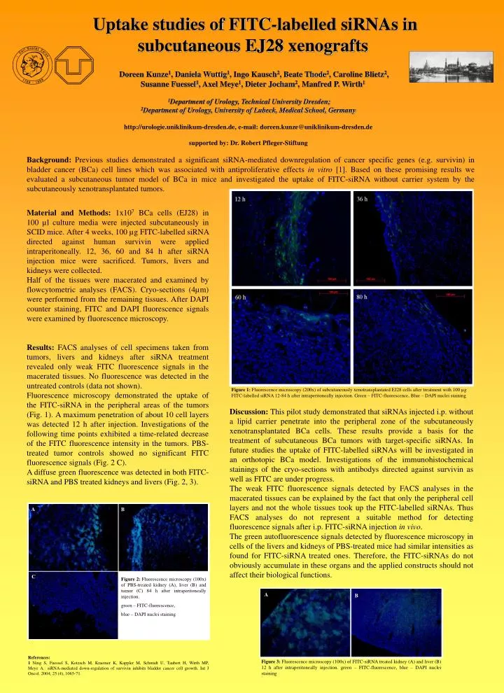
Figure 1: Fluorescence microscopy (200x) of subcutaneously xenotransplantated EJ28 cells after treatment with 100 µg FITC-labelled siRNA 12-84 h after intraperitoneally injection. Green – FITC-fluorescence, Blue – DAPI nuclei staining A B A B C Figure 2:Fluorescence microscopy (100x) of PBS-treated kidney (A), liver (B) and tumor (C) 84 h after intraperitoneally injection. green – FITC-fluorescence, blue – DAPI nuclei staining Figure 3: Fluorescence microscopy (100x) of FITC-siRNA treated kidney (A) and liver (B) 12 h after intraperitoneally injection. green – FITC-fluorescence, blue – DAPI nuclei staining • Uptake studies of FITC-labelled siRNAs in • subcutaneous EJ28 xenografts • Doreen Kunze1, Daniela Wuttig1, Ingo Kausch2, Beate Thode2, Caroline Blietz2, • Susanne Fuessel1, Axel Meye1, Dieter Jocham2, Manfred P. Wirth1 • 1Department of Urology, Technical University Dresden; • 2Department of Urology, University of Lubeck, Medical School, Germany • http://urologie.uniklinikum-dresden.de, e-mail: doreen.kunze@uniklinikum-dresden.de • supported by: Dr. Robert Pfleger-Stiftung Background: Previous studies demonstrated a significant siRNA-mediated downregulation of cancer specific genes (e.g. survivin) in bladder cancer (BCa) cell lines which was associated with antiproliferative effects in vitro [1]. Based on these promising results we evaluated a subcutaneous tumor model of BCa in mice and investigated the uptake of FITC-siRNA without carrier system by the subcutaneously xenotransplantated tumors. Material and Methods: 1x10⁷ BCa cells (EJ28) in 100 µl culture media were injected subcutaneously in SCID mice. After 4 weeks, 100 µg FITC-labelled siRNA directed against human survivin were applied intraperitoneally. 12, 36, 60 and 84 h after siRNA injection mice were sacrificed. Tumors, livers and kidneys were collected. Half of the tissues were macerated and examined by flowcytometric analyses (FACS). Cryo-sections (4µm) were performed from the remaining tissues. After DAPI counter staining, FITC and DAPI fluorescence signals were examined by fluorescence microscopy. Results:FACS analyses of cell specimens taken from tumors, livers and kidneys after siRNA treatment revealed only weak FITC fluorescence signals in the macerated tissues. No fluorescence was detected in the untreated controls (data not shown). Fluorescence microscopy demonstrated the uptake of the FITC-siRNA in the peripheral areas of the tumors (Fig. 1). A maximum penetration of about 10 cell layers was detected 12 h after injection. Investigations of the following time points exhibited a time-related decrease of the FITC fluorescence intensity in the tumors. PBS-treated tumor controls showed no significant FITC fluorescence signals (Fig. 2 C). A diffuse green fluorescence was detected in both FITC-siRNA and PBS treated kidneys and livers (Fig. 2, 3). Discussion: This pilot study demonstrated that siRNAs injected i.p. without a lipid carrier penetrate into the peripheral zone of the subcutaneously xenotransplantated BCa cells. These results provide a basis for the treatment of subcutaneous BCa tumors with target-specific siRNAs. In future studies the uptake of FITC-labelled siRNAs will be investigated in an orthotopic BCa model.Investigations of the immunohistochemical stainings of the cryo-sections with antibodys directed against survivin as well as FITC are under progress. The weak FITC fluorescence signals detected by FACS analyses in the macerated tissues can be explained by the fact that only the peripheral cell layers and not the whole tissues took up the FITC-labelled siRNAs. Thus FACS analyses do not represent a suitable method for detecting fluorescence signals after i.p. FITC-siRNA injection in vivo. The green autofluorescence signals detected by fluorescence microscopy in cells of the livers and kidneys of PBS-treated mice had similar intensities as found for FITC-siRNA treated ones. Therefore, the FITC-siRNAs do not obviously accumulate in these organs and the applied constructs should not affect their biological functions. References: 1 Ning S, Fuessel S, Kotzsch M, Kraemer K, Kappler M, Schmidt U, Taubert H, Wirth MP, Meye A.: siRNA-mediated down-regulation of survivin inhibits bladder cancer cell growth. Int J Oncol. 2004; 25 (4), 1065-71.