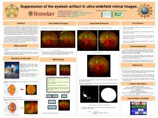
Download
1 / 26
260 likes | 420 Views
ULTRA-FAST LOCALIZATION OF THE OPTIC DISC IN RETINAL FUNDUS IMAGES. Ahmed E. Mahfouz Dr. Ahmed S. Fahmy. Objectives. Project Goal: Automatic Quantitative Analysis of Retinal Images To achieve this goal, localization of retinal anatomical structures is required as an initial step

E N D
ULTRA-FAST LOCALIZATION OF THE OPTIC DISC IN RETINAL FUNDUS IMAGES • Ahmed E. Mahfouz • Dr. Ahmed S. Fahmy
Objectives Project Goal: Automatic Quantitative Analysis of Retinal Images • To achieve this goal, localization of retinal anatomical structures is required as an initial step • In this work, we propose an ultrafast localization of the Optic Disc (OD)
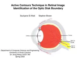
Optic Disc (OD) in Retina Images Vessels Tree Fovea Optic Disc (OD) Colored Retina Fundus Image, STARE Database
Importance of OD localization Localization of the Optic Disc (OD) is useful for: • OD segmentation • Locating other structures (e.g. fovea) • Retinal image registration • Identification of left & right eyes • Tracking retinal vessels
OD Localization: Simple Threshold Retina Fundus Image, STARE Database
OD Localization: Simple Threshold Retina Fundus Image, STARE Database
OD Localization: sophisticated methods Several OD localization methods are available: • Vasculature convergence A. Hoover et al. (2003), A. Youssif et al. (2008) • Geometrical model fitting M. Foracchia et al. (2004) • Template matching M. Lalonde et al. (2003) • Hough Transform F. terHaar (2005)
Vasculature Convergence Original Image Contrast Enhancement Histogram Equalization 13.5 min Final Result Vessels Extraction Thinning 1Aliaa Youssif, AtefGhalwash, and AmrGhoneim "Optic Disc Detection from Normalized digital Fundus Images by means of a Vessels' Direction Matched Filter" IEEE Trans. Medical Imaging, vol.27, no.1, pp. 11–18, Jan. 2008.
Geometric Model Fitting Original Image Contrast Enhancement Histogram Equalization 22 min Final Result Vessels Extraction Model Fitting • 2M. Foracchia, E. Grisan, A. Ruggeri, "Detection of Optic Disc in Retinal Images by Means of a Geometrical Model of Vessel Structure," IEEE Trans. Medical Imaging, vol. 23, no. 10, pp. 1189 – 1195, Oct. 2004.
Motivation • Although available techniques have high success rate, they are impractical due to their long computation times. • OD localization is a preprocessing stepthat provides a seed for further processing steps (segmentation, registration, …etc) New fast localization techniques are required
Ultra-Fast Localization • Search Space Dimensionality Reduction ×
Ultra-Fast Localization Projected Features: Difference between vertical and horizontal edges (MAX) Pixels’ intensity (MIN) Horizontal Localization • Window height = image height • Window width = 2 X main vessel thickness Projection of the image features on the horizontal-axis
Ultra-Fast Localization Vertical Localization Projection of the image features on the vertical-axis Projected Features: Summation of vertical and horizontal edges (MAX) Pixels’ intensity (MAX)
Ultra-Fast Localization • Directionality of the vessels is described by their corresponding edges • No matched filters are used for vessels extraction • The horizontal and vertical edge maps are acquired by applying a 3 X 1 gradient mask [1 0 -1] and its transpose to the retinal image
Evaluation • The new technique is evaluated using four publicly available databases (mostly used in literature): STARE,DRIVE, DIARETDB0, DIARETDB1 • The new technique correctly located the OD in 321 images out of 340 images (94.4% accuracy) • The new technique achieved a computation time of 0.2 – 0.7 seconds depending on the image resolution
Results (failure) This work has been accepted for presentation in 2009 IEEE Int. Conf. on Image Processing (ICIP’09)
Accuracy Improvement • In the previous technique, only the following image features have been used: • Edge information of the retinal vessels. • Pixels’ Intensity • In order to improve the accuracy of the technique, we propose using additional image features such as “Geometric Shape of the Optic Disc”
Accuracy Improvement • Construct a set of candidate locations • 2 candidates • Define a scoring index for each candidate • Index = Value of cand_H at this location • Weight each index with a factor • Geometrical features cand_H
Evaluation Comparing the performance the proposed technique with/without the improvement step on the 4 databases (340 images) This work has been accepted for presentation in the Int. Conf. of Medical Image Computing and Computer Assisted Intervention (MICCAI’09)
Conclusion • A novel technique for fast and accurate OD localization is developed. • The technique is based on reducing the search space dimensionality by projecting image features onto orthogonal axes. • The new technique is much faster (fraction of a second) than currently available techniques (few minutes)
Next Step • Apply the new technique to FluorescienAngiograms (FA) of the retina (high contrast images) • Use the new idea of projecting the image features in other applications, such as: • Registration of retinal images • Determining the orientation of the retinal images (left or right eye)