1 / 14

A 图:窦性心律,正常心电图; B 图: Ⅰ , Ⅱ , Ⅲ , aVF , V2~6 导联 ST 段水平形下降, V5 导联 ST 段下降达 0.25mV , aVR 导联 ST 段抬高。

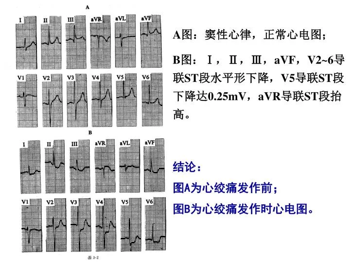
A 图:窦性心律,正常心电图; B 图: Ⅰ , Ⅱ , Ⅲ , aVF , V2~6 导联 ST 段水平形下降, V5 导联 ST 段下降达 0.25mV , aVR 导联 ST 段抬高。. 结论: 图 A 为心绞痛发作前; 图 B 为心绞痛发作时心电图。. 心绞痛发作时心电图. 结论: 图 A 为心绞痛发作前 图 B 为心绞痛发作时心电图. 诊断:慢性冠状动脉供血不足. 诊断:左室肥厚;慢性冠状动脉供血不足。. 急性下壁心肌梗死演变过程. 广泛前壁心肌梗死. 诊断:窦性心律,急性前间壁心肌梗死。.

brody
Download Presentation

A 图:窦性心律,正常心电图; B 图: Ⅰ , Ⅱ , Ⅲ , aVF , V2~6 导联 ST 段水平形下降, V5 导联 ST 段下降达 0.25mV , aVR 导联 ST 段抬高。

An Image/Link below is provided (as is) to download presentation Download Policy: Content on the Website is provided to you AS IS for your information and personal use and may not be sold / licensed / shared on other websites without getting consent from its author. Content is provided to you AS IS for your information and personal use only. Download presentation by click this link. While downloading, if for some reason you are not able to download a presentation, the publisher may have deleted the file from their server. During download, if you can't get a presentation, the file might be deleted by the publisher.

E N D

Presentation Transcript


  1. A图:窦性心律,正常心电图; B图:Ⅰ,Ⅱ,Ⅲ,aVF,V2~6导联ST段水平形下降,V5导联ST段下降达0.25mV,aVR导联ST段抬高。 结论: 图A为心绞痛发作前; 图B为心绞痛发作时心电图。

  2. 心绞痛发作时心电图

  3. 结论: 图A为心绞痛发作前 图B为心绞痛发作时心电图

  4. 诊断:慢性冠状动脉供血不足

  5. 诊断:左室肥厚;慢性冠状动脉供血不足。

  6. 急性下壁心肌梗死演变过程

  7. 广泛前壁心肌梗死

  8. 诊断:窦性心律,急性前间壁心肌梗死。

  9. Ⅱ,Ⅲ,aVF导联ST段抬高呈单向曲线;Q波时限>0.04秒;Ⅰ,aVL导联ST段下降0.1~0.3mV。Ⅱ,Ⅲ,aVF导联ST段抬高呈单向曲线;Q波时限>0.04秒;Ⅰ,aVL导联ST段下降0.1~0.3mV。 诊断:窦性心律,急性下壁心肌梗死,一度房室传导阻滞。

  10. Ⅰ、aVL导联出现异常Q波,ST段抬高,对应Ⅱ、Ⅲ、aVF导联ST段下降。Ⅰ、aVL导联出现异常Q波,ST段抬高,对应Ⅱ、Ⅲ、aVF导联ST段下降。 诊断:窦性心律,急性高侧壁心肌梗死。

  11. 诊断: 窦性心律; 急性前壁合并下壁心肌梗死。

  12. 诊断: 窦性心律; 急性下壁合并正后壁心肌梗死。

More Related