Download
1 / 75
750 likes | 843 Views
UNIT B. Chapter 12: Nervous System. Section 12.1. The nervous system coordinates and regulates the functioning of the body’s other systems. The nervous system consists of two major systems that work together: Central nervous system (CNS): brain and spinal cord

E N D
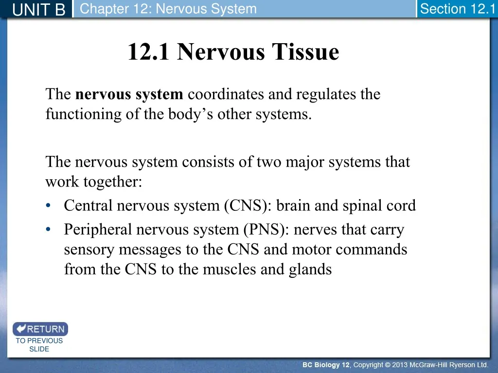
UNIT B Chapter 12: Nervous System Section 12.1 The nervous system coordinates and regulates the functioning of the body’s other systems. The nervous system consists of two major systems that work together: Central nervous system (CNS): brain and spinal cord Peripheral nervous system (PNS): nerves that carry sensory messages to the CNS and motor commands from the CNS to the muscles and glands 12.1 Nervous Tissue TO PREVIOUS SLIDE
UNIT B Chapter 12: Nervous System Section 12.1 Figure 12.1 Organization of the nervous system. The sensory neurons of the peripheral nervous system take nerve impulses from sensory receptors to the central nervous system (CNS), and motor neurons take nerve impulses from the CNS to muscles and glands. TO PREVIOUS SLIDE
UNIT B Chapter 12: Nervous System Section 12.1 The nervous system contains two types of cells: Neurons: cells that transmit nerve impulses between parts of the nervous system Neuroglia: support and nourish neurons, maintain homeostasis, form myelin that surrounds neurons, and aid in signal transmission TO PREVIOUS SLIDE
UNIT B Chapter 12: Nervous System Section 12.1 There are three classes of neurons: Sensoryneurons: take messages to the CNS; have sensory receptors that detect changes in the environment Interneurons: receive input from sensory neurons and other interneurons in the CNS Motorneurons: take messages away from the CNS to an effector (an organ, muscle fibre, or gland); Effectors carry out responses to environmental changes Types of Neurons and Neuron Structure Figure 12.2 Types of neurons. TO PREVIOUS SLIDE
UNIT B Chapter 12: Nervous System Section 12.1 Neurons vary in appearance, but most of them have three parts: Cell body: contains the nucleus and other organelles Dendrites: extensions leading toward the cell body that receive signals from other neurons and send them to the cell body Axon: conducts nerve impulses away from the cell body toward other neurons or effectors Figure 12.2 Types of neurons. TO PREVIOUS SLIDE
UNIT B Chapter 12: Nervous System Section 12.1 Some axons are covered by a protective myelin sheath. In the PNS, a myelin sheath is formed by Schwann cells, a type of neuroglia that contains myelin in the plasma membranes Schwann cells wrap around an axon and lay down many layers of plasma membrane Each Schwann cell myelinates only part of an axon, leaving gaps called nodes of Ranvier Myelin Sheath Figure 12.3 Myelin sheath. a. In the PNS, a myelin sheath forms when Schwann cells wrap themselves around an axon. b. Electron micrograph of a cross section of an axon surrounded by a myelin sheath. TO PREVIOUS SLIDE
UNIT B Chapter 12: Nervous System Section 12.1 Myelin in the PNS The myelin sheath plays an important role in nerve generation in the PNS If an axon is severed, the myelin sheath remains and serves as a passageway for new fibre growth Myelin in the CNS In the CNS, myelin is produced by oligodendrocytes, a type of neuroglia Nerve regeneration does not occur to any significant degree in the CNS TO PREVIOUS SLIDE
UNIT B Chapter 12: Nervous System Section 12.1 Nervous tissue in the CNS The CNS is composed of two types of nervous tissue: Grey matter Contains neurons with short, nonmyelinated axons Found in the surface layer of the brain and the central part of the spinal cord White matter Contains myelinated axons that run together in bundles called tracts Found deep within the grey matter of the brain and surrounds the grey matter in the spinal cord TO PREVIOUS SLIDE
UNIT B Chapter 12: Nervous System Section 12.2 The nervous system uses the nerve impulse to convey information. The nerve impulse can be studied using excised axons and a voltmeter called an oscilloscope. Voltage: measured in millivolts (mV); a measure of the electrical potential difference between two points In a neuron, the two points are in the inside (axoplasm) and the outside of the axons On a voltmeter, voltage is displayed as a trace (pattern) over time 12.2 Transmission of Nerve Impulses TO PREVIOUS SLIDE
UNIT B Chapter 12: Nervous System Section 12.2 In an axon that is not conducting an impulse, the voltmeter records a potential difference across an axon membrane equal to -70mV. This reading, known as the resting potential, shows that the inside of the axon is negative compared to the outside (there is polarity across the axonal membrane) The resting potential is the potential difference across the membrane in a resting neuron Resting Potential TO PREVIOUS SLIDE
UNIT B Chapter 12: Nervous System Section 12.2 The polarity of the resting axonal membrane is due to a difference in ion distribution on each side. The concentration of Na+ is greater outside the axon than inside The concentration of K+ is greater inside the axon than outside Figure 12.4 Action Potential. TO PREVIOUS SLIDE
UNIT B Chapter 12: Nervous System Section 12.2 This unequal distribution is maintained by carrier proteins called sodium-potassium pumps, which actively transport Na+ out of the axon and K+ into the axon The pumps are always working because the membrane is permeable to Na+ and K+ The membrane is more permeable to K+, therefore there are always more positive ions outside the membrane than inside Negatively charged organic ions on the inside of the axon also contribute to the polarity across a resting axonal membrane TO PREVIOUS SLIDE
UNIT B Chapter 12: Nervous System Section 12.2 An action potential is a rapid change in polarity across an axonal membrane as the nerve impulse occurs. An all-or-none phenomenon: if a stimulus causes the membrane to depolarize to a certain level (threshold), an action potential occurs The strength of an action potential does not change, but an intense stimulus can cause an axon to fire (start an action potential) more often Requires two gated channel proteins in the membrane: One channel protein allows Na+ to pass into the axon One channel protein allows K+ to pass out of the axon Action Potential TO PREVIOUS SLIDE
UNIT B Chapter 12: Nervous System Section 12.2 Figure 12.4 Action Potential. c. The changes in the transmembrane potential of the axon are a result of sodium ions flowing into the axon and potassium ions flowing out. An action potential lasts only a few seconds. TO PREVIOUS SLIDE
UNIT B Chapter 12: Nervous System Section 12.2 Sodium Gates Open (Depolarization) When an action potential begins, sodium channel gates open, and Na+ flows down its concentration gradient into the axon As Na+ moves inside the axon, the membrane potential changes from -70 mV to +35 mV This is called depolarization because the charge inside the axon changes from negative to positive Action Potential: Sequence of Events TO PREVIOUS SLIDE
UNIT B Chapter 12: Nervous System Section 12.2 Figure 12.4 Action Potential. a. The action potential begins as the sodium gates (purple) open and Na+ ions move into the axon through facilitated diffusion. This is depolarization as the membrane potential jumps from −70 to +35 millivolts. TO PREVIOUS SLIDE
UNIT B Chapter 12: Nervous System Section 12.2 Potassium Gates Open (Repolarization) The potassium channel gates open, and K+ flows down its concentration gradient out of the axon As K+ flows out of the axon, the action potential becomes more negative again (repolarization) During this time, it briefly becomes slightly more negative that its original resting potential (hyperpolarization) Action Potential: Sequence of Events TO PREVIOUS SLIDE
UNIT B Chapter 12: Nervous System Section 12.2 Figure 12.4 Action Potential. b. The repolarization of a neuron occurs as the potassium gates (orange) open and K+ ions move out of the axon through facilitated diffusion. TO PREVIOUS SLIDE
UNIT B Chapter 12: Nervous System Section 12.2 Action potentials in nonmyelinated axons The action potential travels down an axon one small section at a time When an action potential has moved on, the previous section undergoes a refractory period, during which the sodium gates are unable to open The action potential cannot move backward; it always moves down an axon When the refractory period is over, the sodium-potassium pump has restored the ion distribution by pumping Na+ out of the axon and K+ into the axon Conduction of an Action Potential TO PREVIOUS SLIDE
UNIT B Chapter 12: Nervous System Section 12.2 Action potentials in myelinated axons The gated ion channels that produce an action potential are concentrated at the nodes of Ranvier Ion exchange only occurs at these nodes, therefore the action potential travels faster than in nonmyelinated axons The action potential “jumps” from node to node (saltatory conduction) TO PREVIOUS SLIDE
UNIT B Chapter 12: Nervous System Section 12.2 Every axon branches into endings that have a small swelling called an axon terminal Each terminal lies close to the dendrite or cell body of another neuron or a muscle cell This region of close proximity is called a synapse or chemical synapse Membrane of the first neuron: presynaptic membrane Membrane of the second neuron: postsynaptic membrane Two neurons at a synapse do not physically touch each other; they are separated by a tiny gap called the synaptic cleft Transmission Across a Synapse TO PREVIOUS SLIDE
UNIT B Chapter 12: Nervous System Section 12.2 An action potential cannot cross a synapse. Communication between two neurons at a chemical synapse is carried out by neurotransmitters(chemicals stored in the synaptic vesicles in axon terminals) Figure 12.5 Structure and function of a synapse. Transmission across a synapse from one neuron to another occurs when an action potential causes a neurotransmitter to be released at the presynaptic membrane. TO PREVIOUS SLIDE
UNIT B Chapter 12: Nervous System Section 12.2 When an action potential arrives at an axon terminal: Gated channels for Ca2+ open, and Ca2+ enters the terminal Ca2+ interacts with contractile proteins, which contract and pull the synaptic vesicles to the presynaptic membrane Rise in Ca2+ stimulates synaptic vesicles to merge with the presynapticmembrane, resulting in exocytosis TO PREVIOUS SLIDE
UNIT B Chapter 12: Nervous System Section 12.2 Neurotransmitter molecules are released into the synaptic cleft and diffuse across the synapse to the postsynaptic membrane, where they bind to specific receptors TO PREVIOUS SLIDE
UNIT B Chapter 12: Nervous System Section 12.2 Depending on the neurotransmitter, the postsynaptic neuron can either be excited (causing an action potential) or inhibited (stopping an action potential) TO PREVIOUS SLIDE
UNIT B Chapter 12: Nervous System Section 12.2 A neuron may receive many excitatory and inhibitory signals since its dendrites and cell body can have synapses with many other neurons. Excitatory signals: cause a depolarizing effect Inhibitory signals: cause a hyperpolarizing effect Synaptic Integration Figure 12.6 Synaptic integration. TO PREVIOUS SLIDE
UNIT B Chapter 12: Nervous System Section 12.2 Synaptic integration is the summing up of the excitatory and inhibitory signals in a postsynaptic neuron. If the combined signals cause the membrane potential to rise above threshold, an action potential will occur Figure 12.6 Synaptic integration. a. Inhibitory signals and excitatory signals are summed up in the dendrites and cell body of the postsynaptic neuron. Only if the combined signals cause the membrane potential to rise above threshold does an action potential occur. b. In this example, threshold was not reached. TO PREVIOUS SLIDE
UNIT B Chapter 12: Nervous System Section 12.2 Once a neurotransmitter has been released into a synaptic cleft and has initiated a response, it is removed from the cleft. This prevents continuous stimulation (or inhibition) of postsynaptic membranes In some synapses, the postsynaptic membrane contains enzymes that break down the neurotransmitter In other synapses, the presynaptic membrane reabsorbs the neurotransmitter for repackaging in synaptic vesicles Neurotransmitters TO PREVIOUS SLIDE
UNIT B Chapter 12: Nervous System Section 12.2 Many drugs that affect the nervous system act by interfering or enhancing the action of neurotransmitters. Drugs can: Enhance or block the release of neurotransmitter Mimic the neurotransmitter Block the receptor for the neurotransmitter Interfere with the removal of the neurotransmitter Example: Sarin gas is a chemical weapon that inhibits acetylcholinesterase (AChE), an enzyme that is responsible for the breakdown of acetylcholine (ACh) Leads to prolonged ACh activity (convulsive spasms) Neurotransmitters TO PREVIOUS SLIDE
UNIT B Chapter 12: Nervous System Section 12.3 The central nervous system is composed of the spinal cord and the brain. Brain: controls breathing, heart rate, body temperature, blood pressure, emotions, reasoning, memory, and creativity Spinal cord: a means of communication between the brain and the peripheral nerves that leave the cord 12.3 The Central Nervous System TO PREVIOUS SLIDE
UNIT B Chapter 12: Nervous System Section 12.3 The brain and spinal cord are wrapped in protective membranes called meninges The spaces between meninges are filled with cerebrospinalfluid, which cushions and protects the CNS This fluid is produced and stored in the brain’s ventricles (hollow cavities)and the spinal cord’s central canal If the fluid accumulates in the brain and does not properly drain, the brain can push against the skull, causing brain damage TO PREVIOUS SLIDE
UNIT B Chapter 12: Nervous System Section 12.3 Figure 12.7 Organization of the nervous system. The CNS is composed of the spinal cord and brain. The PNS is composed of the motor and sensory pathways. TO PREVIOUS SLIDE
UNIT B Chapter 12: Nervous System Section 12.3 Structure of the Spinal Cord Individual vertebra protect the spinal cord Spinal nerves project from the cord between the vertebrae in the vertebral column Fluid-filled intervertebraldisks cushion and separate the vertebrae The Spinal Cord Figure 12.8 Spinal cord. a. The spinal cord passes through the vertebral canal formed by the vertebrae. TO PREVIOUS SLIDE
UNIT B Chapter 12: Nervous System Section 12.3 Central canal: contains the cerebrospinal fluid Grey matter: centrally located, shaped like the letter H Contains parts of sensory neurons, motor neurons, and interneurons Dorsal root: contains sensory fibres entering grey matter Ventral root: contains motor fibres exiting grey matter Spinal nerves: part of PNS Figure 12.8 Spinal cord. b. The spinal cord has a central canal filled with cerebrospinal fluid, grey matter in an H-shaped configuration, and white matter. The white matter contains tracts that take nerve impulses to and from the brain. TO PREVIOUS SLIDE
UNIT B Chapter 12: Nervous System Section 12.3 White matter: surrounds grey matter Contains ascending tracts taking information to the brain and descending tracts taking information from the brain Tracts cross each other after entering and exiting CNS Left side of brain: controls right side of body Right side of brain: controls left side of body Figure 12.8 Spinal cord. c. Photomicrograph of a cross section of the spinal cord. TO PREVIOUS SLIDE
UNIT B Chapter 12: Nervous System Section 12.3 Functions of the Spinal Cord The spinal cord sends sensory information to the brain, receives motor input from the brain, and carries out reflex actions. Example: Sensation When someone touches your hand, sensory receptors generate nerve impulses that pass through sensory fibres to the spinal cord and up ascending tracts to the brain Example: Voluntary movement When we move our limbs, motor impulses in the brain pass down descending tracts to the spinal cord and out to our muscles through motor fibres TO PREVIOUS SLIDE
UNIT B Chapter 12: Nervous System Section 12.3 The brain has four major parts: Cerebrum (two lateral ventricles) Diencephalon (third ventricle) Cerebellum (fourth ventricle) Brain stem (fourth ventricle) The Brain TO PREVIOUS SLIDE
UNIT B Chapter 12: Nervous System Section 12.3 Figure 12.9 The human brain. a. The cerebrum, seen here in longitudinal section, is the largest part of the brain in humans. The right cerebral hemisphere is shown here. TO PREVIOUS SLIDE
UNIT B Chapter 12: Nervous System Section 12.3 The cerebrum is the largest part of the brain in humans Communicates with and coordinates activities of other parts of the brain The Cerebrum TO PREVIOUS SLIDE
UNIT B Chapter 12: Nervous System Section 12.3 Structure and Function of the Cerebrum The cerebrum has two halves (cerebralhemispheres) that communicate via the corpus callosum, a bridge of nerve tracts. The cerebral cortex is a thin outer layer of grey matter that covers the cerebral hemispheres Grooves called sulci divide the hemisphere into four lobes: frontal, parietal, occipital, temporal Figure 12.9 The human brain. The cerebrum has left and right cerebral hemispheres, which are connected by the corpus callosum. TO PREVIOUS SLIDE
UNIT B Chapter 12: Nervous System Section 12.3 Figure 12.10 The lobes of a cerebral hemisphere. Each cerebral hemisphere is divided into four lobes: frontal, parietal, temporal, and occipital. The frontal lobe contains centres for reasoning and movement, the parietal lobe for somatic sensing and taste, the temporal lobe for hearing, and the occipital lobe for vision. TO PREVIOUS SLIDE
UNIT B Chapter 12: Nervous System Section 12.3 Frontal Lobe Primary motor area: involved in voluntary movement Premotor area: involved in organizing motor functions Prefrontal area: processing centre involved in reasoning and planning Broca’s area: involved in speech musculature (lips, tongue, larynx) TO PREVIOUS SLIDE
UNIT B Chapter 12: Nervous System Section 12.3 Parietal Lobe Primary somatosensory area: involved in somatic sensing Primary taste area: involved in taste Somatosensory association area: processes and analyzes sensory information from skin and muscles TO PREVIOUS SLIDE
UNIT B Chapter 12: Nervous System Section 12.3 Temporal Lobe Primary auditory area: involved in hearing Auditory association area: associates new audio information with previous audio information Wernicke’sarea: helps us understand written and spoken words TO PREVIOUS SLIDE
UNIT B Chapter 12: Nervous System Section 12.3 Occipital Lobe Primary visual area: involved in vision Visual association area: associates new visual information with previous visual information (e.g., facial recognition) TO PREVIOUS SLIDE
UNIT B Chapter 12: Nervous System Section 12.3 12.11 The primary motor and somatosensory areas. In these drawings, the size of the body part reflects the amount of cerebral cortex devoted to that body part. For example, the amount of primary motor cortex (a) and somatosensory cortex (b) devoted to the thumb, fingers, and hand is greater than that for the foot and toes. TO PREVIOUS SLIDE
UNIT B Chapter 12: Nervous System Section 12.3 Central White Matter Most of the cerebrum beneath the cerebral cortex is composed of white matter Tracts within the cerebrum take information between different sensory, motor, and association areas Basal Nuclei Basalnuclei are masses of grey matter located deep within the white matter of the cerebrum Integrate motor commands to ensure proper muscle groups are activated or inhibited TO PREVIOUS SLIDE
UNIT B Chapter 12: Nervous System Section 12.3 Thediencephalonis a region that encircles the third ventricle. The Diencephalon TO PREVIOUS SLIDE
UNIT B Chapter 12: Nervous System Section 12.3 Structure and Function of the Diencephalon Hypothalamus Integrating centre that helps maintain homeostasis Regulates hunger, sleep, thirst, body temperature, and water balance Controls the pituitary gland and serves as a link between the nervous and endocrine systems TO PREVIOUS SLIDE