Download
1 / 68
710 likes | 956 Views
A Review of Creutzfeldt-Jakob Disease (CJD) with an Emphasis on Clinical Laboratory Issues. Jeannie Druckenmiller, BS, SM(NRCM), CIC Wisconsin Division of Public Health (DPH) June 13, 2012. Objectives.

E N D
A Review of Creutzfeldt-Jakob Disease (CJD) with an Emphasis on Clinical Laboratory Issues Jeannie Druckenmiller, BS, SM(NRCM), CIC Wisconsin Division of Public Health (DPH) June 13, 2012
Objectives • Describe commonly encountered prion diseases including issues pertaining to the incidence, surveillance, transmission, and epidemiology of human prion diseases. • Describe current infection control guidelines for safe handling of specimens suspected of harboring prion disease in the clinical laboratory. • Describe types of tests for prions and appropriate handling and transport of clinical specimens to be tested for CJD.
Incident: 2009 • Hospitalized patient with rapidly progressive dementia and ataxia • Tumor found on brain CT, biopsy done – not malignant; no histology present indicating prion disease • Patient’s mental status continues to rapidly deteriorate; patient dies within weeks • CJD discovered following full brain autopsy • In the interim, the neurosurgical instruments used on the patient had been used on > 50 other patients without special prion decontamination reprocessing
Incident: cont. • Per hospital policy, the biopsy on the tumor was considered to be neurosurgery on a ‘space-occupying lesion,’ hence Infection Prevention was not notified in advance. • Instruments were retrieved and reprocessed using prion protocol • > 50 patients were notified of possible exposure to CJD
Characteristics of Transmissible Spongiform Encephalopathies (TSEs) of Humans • Rare, progressive neurodegenerative disorders • Occur worldwide • Invariably fatal • No treatment • Produce no immune response • Caused by accumulation of abnormal prion protein • Incubation period months to >20 years • Prion extremely resistant to inactivation • Are not select agents
Transmissible Spongiform Encephalopathies (TSEs) of Humans • Kuru • Gertsmann-Straussler-Scheinker Syndrome (GSS) • Fatal Familial Insomnia (FFI) • Creutzfeldt-Jakob Disease (CJD) • Variant CJD (vCJD), “Mad Cow,” 1995
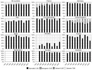
Transmissible Spongiform Encephalopathies (TSEs) of Humans • Kuru – Now eradicated • Gertsmann-Straussler-Scheinker Syndrome (GSS) – Incidence 1:40 million • Fatal Familial Insomnia (FFI) – Incidence 1:40 million • Creutzfeldt-Jakob Disease (CJD) – Incidence 1:1million • Variant CJD (vCJD), “Mad Cow,” 1995 – Over 200 cases reported; most of them in the United Kingdom
Prion Hypothesis PrP res PrP sen • Aberrant form of normal host cellular prion protein: (unfolding and flipping of normal conformation into an abnormal form which is protease resistant) Normal Abnormal • Function of normal cellular prion protein unclear • Cascade of normal form to abnormal configuration
Prion diseases of animals –all caused by distinct prions • Scrapie in sheep and goats • Mink transmissible encephalopathy • Bovine Spongiform Encephalopathy (a.k.a. “mad cow disease”) • Chronic Wasting Disease of deer and elk
New variant CJD • Associated with bovine spongiform encephalopathy (BSE) • vCJD has never been reported in US • Three cases that acquired the disease elsewhere have died in the US • Over 250 cases reported worldwide; most of them in United Kingdom • Association of vCJD with BSE is the first instance of apparent transmission of a TSE across the species barrier Source: Rutala and Weber, 2010.
New variant CJD versus sporadic CJD vCJD sCJD Longer clin. course (12-15 mo) Shorter course (~4-7 mo) More predominant psychiatric sx Earlier onset of dementia; (psychosis, depression, anxiety) myoclonus more pronounced Absence of characteristic EEG Characteristic periodic EEG changes complexes Widespread, numerous amyloid Relative lack of amyloid plaques (florid) in cerebellum plaques typically in cerebral and cerebrum cortex only Geographic and temporal No such correlation with BSEcorrelation with BSE
Normal degradation of PrPsen PrP sen Endocytosis & lysosomal degradation Golgi apparatus
PrP res No lysosomal degradation Cellular disruption from PrPres PrPres accumulates in neural cells disrupting function, leading to vacuolization and cell death
DPH Surveillance of Human Prion Disease:Impetus • Report of first cases of vCJD in U.K. (1995) • First finding of CWD in Wisconsin deer (2002) • Media reports of apparent “CJD cluster” in NW Wisconsin – 2002 • Reports of BSE in North American cattle
Prion disease surveillance is doneby WI Division of Public Health (DPH) - Madison • Prion disease reports should go directly to State DPH • If reported to a Local Health Dept. (LHD), no follow up is required other than to forward the report to DPH • Call Jeannie Druckenmiller at 608-516-5847 or Jim Kazmierczak at 608-266-2154 • Prefer phone reports because time is of the essence(suspect cases ideally identified ante mortem)
Reporting requirement • In Wisconsin, TSEs are reportable directly to the state epidemiolgist (not to local health department (LHD) • Wis Stats Chapters 250 and 252 • Administrative Rule 145.04(4) • Federal Health Insurance Portability and Accountability Act (HIPAA) – Privacy rule: [42 USCA Section 1320(b)] and [45 CFR Section 164.512(b)(1)(i)].
WI DPH Surveillance Activities • Case Finding: - Ongoing review of death certificates - Reporting by clinicians, Infection Control, LHDs - All CSF test results for Tau & 14-3-3 done at NPDPSC are forwarded to DPH • Chart review and basic risk factor information is collected on every potential case • Autopsies on suspect cases encouraged; - funding available for autopsies and transport of remains - specimens sent to NPDPSC • Maintain registry of persons known to have consumed venison from CWD positive deer for later comparison to CJD case list
Summary of Wisconsin CJD Surveillance, 1997 -2011 69 sporadic CJD, 2 familial CJD
Drs. Creutzfelt and Jakob first reported the disease in 1920-1921
CJD – Clinical Signs & Symptoms • Prodrome of confusion, weakness, personality changes, bizarre behavior • Confusion • Dementia – rapidly progressing • Myoclonus – jerking movements • Ataxia • Visual disturbances (visual field cuts, cortical blindness)
CJD – Clinical Characteristics • No fever • No systemic features • Most lab test results are normal • Infectivity of central nervous system tissues persists during and throughout illness
Diagnosis of CJD • Increased 14-3-3 and tau protein in CSF • Rapidly progressive dementia with ataxia • Characteristic brain MRI • Characteristic EEG • Rule out of other neurological diseases and syndromes • Brain biopsy ??? • Diagnosis usually made from brain autopsy
Diagnosis of CJD • Definitive diagnosis can only be done by brain biopsy or post-mortem examination of brain tissue - Histopathology - Immunohistochemical (IHC) staining - Western blot analysis
Lab tests available for CJD: 14-3-3 protein • CSF 14-3-3 protein is sometimes increased in suspect patients • Results reported as negative, positive or ambiguous • Good sensitivity (negative test has high probability of being a true negative) • Significantly affected by presence of blood in the specimen or inflammation in the brain • Test recommended by World Health Organization (WHO) as the screening test
Lab tests available for CJD: Tau protein • Done by National Prion Disease Pathology Surveillance Center (NPDPSC), Cleveland, OH • Has fewer false positives • Reported as a numeric value with a decision point and accompanying positive predictive value • Much less affected by blood in the specimen • The Tau and 14-3-3 tests are complimentary
Lab tests available for CJD • All major reference labs except Mayo send specimens to NPDPSC for Tau and 14-3-3 testing. • Mayo does its own 14-3-3 testing and also performs a Neuron Specific Enolase test (NSE). • Most reference labs and hospital labs will send a CSF to NPDPSC on special request. The issue becomes the cost.
CSF Specimen Transport • To the lab - per hospital policy or reference lab policy. Often, other tests will be performed on the specimen. Prions are not fragile; Standard Precautions should be used in specimen handling. • Transport of a specimen to the NPDPSC: • Frozen and shipped on dry ice • Category B shipping regulations for bio-substances apply. These are available on the Internet. • NPDPSC supplies shipping boxes for autopsy tissue (brain).
How does one get a prion disease? • 85-90% appear spontaneously (sCJD) • Familial forms • Ingestion (e.g., Kuru) • Iatrogenic • Dura mater allografts • Corneal transplants • Human growth hormone • Contaminated surgical instruments
Iatrogenic Transmission of CJD • Over 250 cases worldwide • Primarily linked to cadaveric growth hormone (>130), dura mater (>110) and corneal grafts (3) • Six cases linked to contaminated equipment; four associated with neurosurgical instruments and two with EEG depth electrodes. Source: CDC / Rutala & Weber 2010.
Iatrogenic Transmission of CJD • No iatrogenic cases reported since 1976 • Since 1985, human growth hormone has been manufactured by recombinant DNA technology • No cases associated with exposure to the environment • All cases associated with exposure to brain, spinal cord, pituitary, or deep eye tissue.
Transmissionof Prion Diseases • Not spread by direct or indirect contact, droplets or respiratory secretions (airborne) • Not spread by the environment • Experimentally-all TSEs are transmissible to animals, including the inherited forms • Patient care involves use of Standard Precautions only • No isolation necessary
Risk of CJD Transmission Source: CDC; Rutala and Weber
Infection control guidelines regarding disinfection and sterilization of prions: Pertinent Documents • World Health Organization (WHO) Guideline, 1999. • Rutala and Weber, Guideline, 2010.
Infection control guidelines regarding disinfection and sterilization of prions • World Health Organization (WHO) Infection Control Guidelines for Transmissible Spongiform Encephalopathies • Consensus from an international meeting of experts held in Geneva, Switzerland,1999. • Emphasis for disinfection relies on immersion of instrument in 1N NaOH and autoclaving, then cleaning, then subject to routine sterilization.
WHO Document • The vast majority of diagnostic examinations in clinical labs are performed on blood, serum and other blood derivatives, usually with automated analyzing equipment. • “…and strongly recommend that blood specimens from patients with CJD not be considered infectious, and that no special precautions are needed for its handling in clinical laboratories.”
WHO Document • Similarly, except for CSF, other body fluids, secretions and excretions contain no infectivity and need no special handling.
WHO Document • CSF from patients with CJD • May be infectious • Recommend analysis not be performed in automated equipment • Materials coming in contact with the CSF must either be incinerated or decontaminated according to WHO protocol.
WHO Document • General recommendations include • No eating, drinking, smoking or food in the lab • Use of personal protective equipment (PPE) • Use of disposable equipment whenever possible • Work surface decontamination • Prion contaminated materials be discarded by incineration
WHO Document • Exposure to intact skin • Wash with water • Brief exposure (1 min) with 0.1N NaOH or 1:10 bleach • Percutaneous exposure • Encourage bleeding • Wash with warm soapy water • Eye or mucous membrane • Irrigate with copious amounts of tap water or saline • Usefulness of these strategies is unknown
WHO Document • No special decontamination of work surfaces contaminated with low risk tissues • Work surfaces contaminated with high risk tissue • Use NaOH protocol
WHO Document • Waste disposal • TSE infectious waste applies to low and high infectivity tissues • Recommend placing in leak-proof containers and incineration
Infection control guidelines regarding disinfection and sterilization of prions WHO Guideline endorsed / supported by: • Centers for Disease Control and Prevention (CDC) • National Institutes of Health (NIH) • National Prion Disease Pathology Surveillance Center
Infection control guidelines regarding disinfection and sterilization of prions • William A. Rutala, PhD, MPH and David J. Weber, MD, MPH • Guideline for Disinfection and Sterilization of Prion-Contaminated Medical Instruments • Published in Infection Control and Hospital Epidemiology. 2010;31:107-115. • This is the official journal of the Society for Healthcare Epidemiology of America (SHEA)
Rutala and Weber Guideline • Emphasis on • Keeping instruments wet until they can be re-processed • Pre-cleaning/decontamination in washer-disinfector before autoclaving • After the device is clean it should be sterilized by either autoclaving or using a combination of sodium hydroxide (NaOH) and autoclaving.
Rutala and Weber Guideline Emphasis on: • It is essential that with any sterilization process, and especially when prion contamination may be an issue, that the instrument be fully accessible to the sterilant.
Rutala and Weber Guideline - Premises • Patient’s risk of having a prion disease • High risk patient = exhibiting clinical signs and symptoms of CJD • Comparative infectivity of tissue • High risk tissue = brain, spinal cord, posterior eye
Rutala and Weber Guideline - Premises • No known cases of prion disease transmitted by contaminated medical instruments in past several decades • Transmission is inefficient and current cleaning and disinfection methods, “though suboptimal may be preventing disease.”
Rutala and Weber Guideline – Premises • Studies of iatrogenic-associated CJD from 1952-1976 are missing important details regarding methodology of reprocessing. • Did not incorporate a cleaning step – cleaning can reduce microbial load 4-6 log10
Rutala and Weber Guideline – Premises • SHEA guideline is predicated on epidemiological (evidence-based) studies. Other studies have been based on inactivation studies using lumps of tissue.