Download
1 / 50
510 likes | 715 Views
Chapter 16 The Special Senses. Smell, taste, vision, hearing and equilibrium Housed in complex sensory organs Ophthalmology is science of the eye Otolaryngology is science of the ear. Chemical Senses. Interaction of molecules with receptor cells Olfaction (smell) and gustation (taste)

E N D
Chapter 16The Special Senses • Smell, taste, vision, hearing and equilibrium • Housed in complex sensory organs • Ophthalmology is science of the eye • Otolaryngology is science of the ear
Chemical Senses • Interaction of molecules with receptor cells • Olfaction (smell) and gustation (taste) • Both project to cerebral cortex & limbic system • evokes strong emotional reactions
Olfactory Epithelium • 1 square inch of membrane holding 10-100 million receptors • Covers superior nasal cavity and cribriform plate • 3 types of receptor cells
Olfaction: Sense of Smell • Odorants bind to receptors • Na+ channels open • Depolarization occurs • Nerve impulse is triggered
Adaptation & Odor Thresholds • Adaptation = decreasing sensitivity • Olfactory adaptation is rapid • 50% in 1 second • complete in 1 minute • Low threshold • only a few molecules need to be present • methyl mercaptan added to natural gas as warning
Gustatory Sensation: Taste • Taste requires dissolving of substances • Four classes of stimuli--sour, bitter, sweet, and salty • 10,000 taste buds found on tongue, soft palate & larynx
Anatomy of Taste Buds • An oval body consisting of 50 receptor cells surrounded by supporting cells • A single gustatory hair projects upward through the taste pore • Basal cells develop into new receptor cells every 10 days.
Physiology of Taste • Complete adaptation in 1 to 5 minutes • Thresholds for tastes vary among the 4 primary tastes • most sensitive to bitter (poisons) • least sensitive to salty and sweet
Accessory Structures of Eye • Eyelids or palpebrae • protect & lubricate • Tarsal glands • oily secretions keep lids from sticking together • Conjunctiva • stops at corneal edge • dilated BV--bloodshot
Eyelashes & Eyebrows Eyeball = 1 inch diameter 5/6 of Eyeball inside orbit & protected • Eyelashes & eyebrows help protect from foreign objects, perspiration & sunlight • Sebaceous glands are found at base of eyelashes (sty) • Palpebral fissure is gap between the eyelids
Lacrimal Apparatus • About 1 ml of tears produced per day. Spread over eye by blinking. Contains bactericidal enzyme called lysozyme.
Extraocular Muscles • Six muscles that insert on the exterior surface of the eyeball • . • 4 rectus muscles -- superior, inferior, lateral and medial • 2 oblique muscles -- inferior and superior
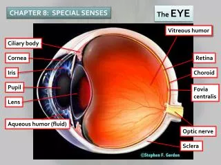
Tunics (Layers) of Eyeball • Fibrous Tunic(outer layer) • Vascular Tunic (middle layer) • Nervous Tunic(inner layer)
Fibrous Tunic -- Description of Cornea • Transparent • Helps focus light(refraction) • astigmatism • Transplants • common & successful • no blood vessels so no antibodies to cause rejection
Fibrous Tunic -- Description of Sclera • “White” of the eye • Dense irregular connective tissue layer -- collagen & fibroblasts • Provides shape & support
Vascular Tunic -- Choroid & Ciliary Body • Choroid • pigmented epithilial cells (melanocytes) & blood vessels • provides nutrients to retina • black pigment in melanocytes absorb scattered light • Ciliary body • ciliary processes • folds on ciliary body • secrete aqueous humor • ciliary muscle • smooth muscle that alters shape of lens
Vascular Tunic -- Iris & Pupil • Colored portion of eye • Shape of flat donut suspended between cornea & lens • Hole in center is pupil • Function is to regulate amount of light entering eye
Vascular Tunic -- Description of lens • Avascular • Crystallin proteins arranged like layers in onion • Clear capsule & perfectly transparent
Nervous Tunic -- Retina • Posterior 3/4 of eyeball • Optic disc • optic nerve exiting back of eyeball • Central retina BV • fan out to supply nourishment to retina • visible for inspection • hypertension & diabetes • Detached retina • trauma (boxing) • fluid between layers • distortion or blindness View with Ophthalmoscope
Rods & Cones--Photoreceptors • Rods----rod shaped • shades of gray in dim light • 120 million rod cells • discriminates shapes & movements • distributed along periphery • Cones----cone shaped • sharp, color vision • 6 million • fovea of macula lutea • densely packed region • at exact visual axis of eye • 2nd cells do not cover cones • sharpest resolution or acuity
Pathway of Nerve Signal in Retina • Light penetrates retina • Rods & cones transduce light into action potentials • Rods & cones excite bipolar cells • Bipolars excite ganglion cells • Axons of ganglion cells form optic nerve leaving the eyeball (blind spot) • To thalamus & then the primary visual cortex
Aqueous Humor • Continuously produced by ciliary body • Flows from posterior chamberinto anterior through the pupil • Glaucoma • increased intraocular pressure that could produce blindness • problem with drainage of aqueous humor
Major Processes of Image Formation • Refraction of light • by cornea & lens • light rays must fall upon the retina • Accommodation of the lens • changing shape of lens so that light is focused • Constriction of the pupil • less light enters the eye
Definition of Refraction • Bending of light as it passes from one substance (air) into a 2nd substance with a different density(cornea) • In the eye, light is refracted by the anterior & posterior surfaces of the cornea and the lens
Refraction by the Cornea & Lens • Image focused on retina is inverted & reversed from left to right • Brain learns to work with that information • 75% of Refraction is done by cornea -- rest is done by the lens
Near Point of Vision and Presbyopia • Near point is the closest distance from the eye an object can be & still be in clear focus • 4 inches in a young adult • 8 inches in a 40 year old • lens has become less elastic • 31 inches in a 60 to 80 year old • Reading glasses may be needed by age 40 • presbyopia • glasses replace refraction previously provided by increased curvature of the relaxed, youthful lens
Correction for Refraction Problems • Emmetropic eye (normal) • can refract light from 20 ft away • Myopia (nearsighted) • eyeball is too long from front to back • glasses concave • Hypermetropic (farsighted) • eyeball is too short • glasses convex (coke-bottle) • Astigmatism • corneal surface wavy • parts of image out of focus
Constriction of the Pupil • Constrictor pupillae muscle contracts • Prevents light rays from entering the eye through the edge of the lens • Sharpens vision by preventing blurry edges • Protects retina very excessively bright light
Convergence of the Eyes • Binocular vision in humans has both eyes looking at the same object • As you look at an object close to your face, both eyeballs must turn inward. • convergence
Photoreceptors • Photopigment is integral membrane protein of outer segment membrane • photopigment membrane folded into “discs” & replaced at a very rapid rate • Photopigments = opsin (protein) + retinal (derivative of vitamin A) • rods contain rhodopsin • cone photopigments contain 3 different opsin proteins permitting the absorption of 3 different wavelengths (colors) of light
Color Blindness & Night Blindness • Color blindness • inability to distinguish between certain colors • absence of certain cone photopigments • red-green color blind person can not tell red from green • Night blindness (nyctalopia) • difficulty seeing in low light • inability to make normal amount of rhodopsin • possibly due to deficiency of vitamin A
Light and Dark Adaptation • Light adaptation • adjustments when emerge from the dark into the light • Dark adaptation • adjustments when enter the dark from a bright situation • light sensitivity increases as photopigments regenerate • during first 8 minutes of dark adaptation, only cone pigments are regenerated, so threshold burst of light is seen as color • after sufficient time, sensitivity will increase so that a flash of a single photon of light will be seen as gray-white
synapse in thalamus & visual cortex Brain Pathways of Vision
Processing of Image Data in the Brain • Visual information in optic nerve travels to • occipital lobe for vision • midbrain for controlling pupil size & coordination of head and eye movements • hypothalamus to establish sleep patterns based upon circadian rhythms of light and darkness
Visual fields • Left occipital lobe receives visual images from right side of an object through impulses from nasal 1/2 of the right eye and temporal 1/2 of the left eye • Left occipital lobe sees right 1/2 of the world • Fibers from nasal 1/2 of each retina cross in optic chiasm
External Ear • Function = collect sounds • Structures • auricle or pinna • elastic cartilage covered with skin • external auditory canal • curved 1” tube of cartilage & bone leading into temporal bone • ceruminous glands produce cerumen = ear wax • tympanic membrane or eardrum • epidermis, collagen & elastic fibers, simple cuboidal epith. • Perforated eardrum (hole is present) • at time of injury (pain, ringing, hearing loss, dizziness) • caused by explosion, scuba diving, or ear infection
Middle Ear Cavity • Air filled cavity in the temporal bone • Separated from external ear by eardrum and from internal ear by oval & round window • 3 ear ossicles connected by synovial joints • malleus attached to eardrum, incus & stapes attached by foot plate to membrane of oval window • stapedius and tensor tympani muscles attach to ossicles • Auditory tube leads to nasopharynx • helps to equalize pressure on both sides of eardrum • Connection to mastoid bone =mastoiditis
Inner Ear---Bony Labyrinth Vestibule canals • Bony labyrinth = set of tubelike cavities in temporal bone • semicircular canals, vestibule & cochlea lined with periosteum & filled with perilymph • surrounds & protects Membranous Labyrinth ampulla
Inner Ear---Membranous Labyrinth • Membranous labyrinth = set of membranous tubes containing sensory receptors for hearing & balance and filled with endolymph • utricle, saccule, ampulla, 3 semicircular ducts & cochlea
Anatomy of the Organ of Corti • 16,000 hair cells have 30-100 stereocilia(microvilli ) • Microvilli make contact with tectorial membrane (gelatinous membrane that overlaps the spiral organ of Corti) • Basal sides of inner hair cells synapse with 1st order sensory neurons whose cell body is in spiral ganglion
Vibrating object causes compression of air around it = sound waves audible range is 20 to 20,000 Hz hear best within 500 to 5000 cycles/sec or Hz speech is 100 to 3000 Hz Frequency of a sound vibration is pitch higher frequency is higher pitch Greater intensity (size) of vibration, the louder the sound measured in decibels (dB) Conversation is 60 dB; pain above 140dB OSA requires ear protection above 90dB Sound Waves
Deafness • Nerve deafness • damage to hair cells from antibiotics, high pitched sounds, anticancer drugs • the louder the sound the quicker the hearing loss • fail to notice until difficulty with speech • Conduction deafness • perforated eardrum • otosclerosis
Physiology of Hearing • Auricle collects sound waves • Eardrum vibrates • slow vibration in response to low-pitched sounds • rapid vibration in response to high-pitched sounds • Ossicles vibrate since malleus attached to eardrum • Stapes pushes on oval window producing fluid pressure waves in scala vestibuli & tympani • oval window vibration 20X more vigorous than eardrum • Pressure fluctuations inside cochlear duct move the hair cells against the tectorial membrane • Microvilli are bent producing receptor potentials
Cochlear Implants • If deafness is due to destruction of hair cells • Microphone, microprocessor & electrodes translate sounds into electric stimulation of the vestibulocochlear nerve • artificially induced nerve signals follow normal pathways to brain • Provides only a crude representation of sounds
Physiology of Equilibrium (Balance) • Static equilibrium • maintain the position of the body (head) relative to the force of gravity • Dynamic equilibrium • maintain body position (head) during sudden movement of any type--rotation, deceleration or acceleration
Detection of Position of Head • Movement of stereocilia or kinocilium results in the release of neurotransmitter onto the vestibular branches of the vestibulocochler nerve
Detection of Rotational Movement • When head moves, the attached semicircular ducts and hair cells move with it • Nerve signals to the brain are generated indicating which direction the head has been rotated