Download
1 / 36
410 likes | 1.02k Views
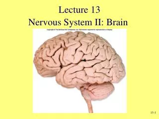
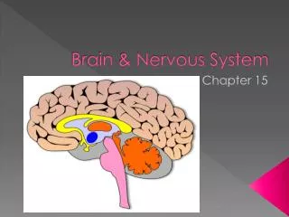
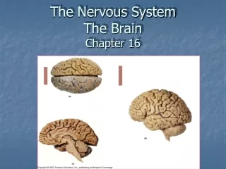
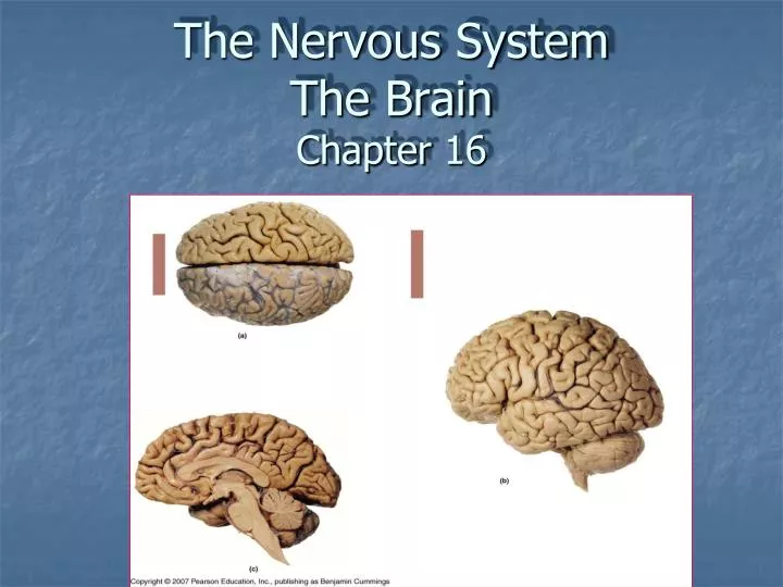
The Nervous System The Brain Chapter 16. The Brain - Overview. Brain stem medulla oblongata (M.O.) pons midbrain (mesencephalon) Diencephalon thalamus hypothalamus epithalamus (pineal gland) Cerebrum Cerebellum. Cerebrum. T. P. P. H. midbrain. Cerebellum. pons. m.o.

E N D
The Brain - Overview • Brain stem • medulla oblongata (M.O.) • pons • midbrain (mesencephalon) • Diencephalon • thalamus • hypothalamus • epithalamus (pineal gland) • Cerebrum • Cerebellum Cerebrum T P P H midbrain Cerebellum pons m.o.
Cranial Meninges Three layers: Dura mater, Arachnoid mater, Pia mater
Dura mater – tough, fibrous outer layer; 2 layers thick around brain (superficial “periosteal layer”/deeper “meningeal layer”) with creation of dural (venous) sinuses between layers, and dural folds into cranial cavity
Falx cerebri Tentorium cerebelli Dural folds • Folds that create septa to subdivide cranial cavity and stabilize the brain. Includes: • falx cerebri – between cerebral hemispheres in longitudinal fissure • tentorium cerebelli – between cerebrum & cerebellum in transverse fissure • falx cerebelli – between cerebellar hemispheres • diaphragma sellae – lines sella turcica
Dural sinuses Superior sagittal sinus Falx cerebri Inferior sagittal sinus Tentorium cerebelli Straight sinus Confluence of sinuses Transverse sinus Sigmoid sinus Spaces between dural layers and dural folds functioning as veins for drainage of blood from cerebral veins, and CSF from subarachnoid space (superor sagittal sinus)
Arachnoid mater– “spidery” web-like middle layer with fine collagen & elastic connections to underlying Pia Mater • Pia mater– delicate, thin inner layer
Subarachnoid space– between arachnoid & pia mater; contains cerebrospinal fluid (CSF) Arachnoid granulations (villi) – projections of arachnoid into dural sinuses for drainage of CSF
Cerebrospinal Fluid (CSF) • clear, colorless fluid formed by filtration of blood plasma by choroid plexuses within ventricles of the brain. • functions in protection of CNS, support, nutrient supply, waste removal
CSF Circulation Lateral ventricles (in cerebral hemispheres) interventricular foramen third ventricle (in diencephalon around and between R/L thalamus) cerebral (mesencephalic) aqueduct of midbrain fourth ventricle (between pons/cerebellum) subarachnoid space & central canal of SC
Reabsorption of CSF through arachnoid granulations (arachnoid villi) of dural sinuses (superior sagittal sinus) into cerebral veins
Blood supply to Brain • Brain requires large amounts of O2 and nutrients (glucose) • Internal carotid arteries + basilar artery (from vertebral arteries) “cerebral arterial circle (of Willis)” • Venous drainage from dural venous sinuses & cerebral veins into vertebral and internal jugular veins
The Brainstem • Medulla oblongata • continuation of the SC above the foramen magnum • contains the pyramidal decussation within the pyramids • cranial nerve nuclei (XII-VIII (cochlear) • cardiac, vasomotor, & respiratory reflex centers • Pons • “bridge” linking cerebellum to SC & other parts of brain via middle cerebellar peduncle • cranial nerve nuclei (VIII (vestibular) – V) • respiratory center
Midbrain (mesencephalon) • cerebral peduncles – location of ascending (sensory) & descending (motor) tracts • tectum – posterior aspect of brainstem; contains • corpora quadrigemina • superior colliculi – visual reflex centers • inferior colliculi – auditory reflex centers • cranial nerve nuclei (IV-III) The Brainstem
The Brainstem • Midbrain (mesencephalon) • substantia nigra – nucleus with dark pigmented neurons that regulate motor output of basal nuclei (basal ganglia) of cerebrum • reticular formation – network of interconnected nuclei throughout brainstem responsible for maintaining states of consciousness
The Diencephalon Thalamus • surrounds 3rd ventricle • 2 sides (left & right thalamus) usually connected by intermediate mass (interthalamic adhesion) • comprised of nuclei that function primarily as sensory relay stations
The Diencephalon • Hypothalamus • connects to pituitary gland via the infundibulum • has many important functions relating to maintaining homeostasis including (but not limited to): -integrating nervous & endocrine systems through control over pituitary gland -integration of ANS from visceral stimuli -hunger/satiety, thirst, body temp. regulation, circadian rhythms -hormone production (ADH, oxytocin) • mamillary bodies – reflex centers associated with eating, & processing of olfactory sensations
The Diencephalon Epithalamus -Pineal gland • secretes Melatonin which helps regulate day-night cycles (circadian rhythm)
The Cerebellum • Separated from cerebrum by transverse fissure • “Tentorium cerebelli” encloses straight sinus & transverse sinus • Two hemisphereres joined by vermis • Hemispheres have 3 lobes – anterior, posterior & flocculonodular • outer cortex of gray matter folded into “folia” • inner white matter “arbor vitae” Transverse fissure
The Cerebellum • Links to brainstem by cerebellar peduncles • inferior cerebellar peduncle M.O. (and S.C.) • middle cerebellar peduncle pons • superior cerebellar peduncle midbrain (and diencephalon/cerebrum) • Functions include: • control of skeletal muscles (unconscious) for balance, coordination & posture • stores patterns of movement
convolutions Cerebrum gyrus sulcus Transverse fissure
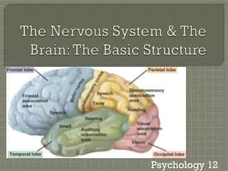
Lateral sulcus (Insula is deep to lateral sulcus) Lobes of Cerebral Hemispheres Central sulcus Parietal lobe Parieto-occipital sulcus (seen along medial surface) Frontal lobe Occipital lobe Temporal lobe
Parieto-occipital sulcus insula
Gray & White matter of cerebrum • Gray matter : • superficial cortex –functional areas includes sensory, motor , & higher order functions • deep cerebral nuclei (aka basal nuclei/basal ganglia) • White matter: • fibers – association commissural projection
White matter of cerebrum Association fibers • association fibers – connect gyri in same hemisphere • commissural fibers – connect gyri in opposite hemispheres (e.g. corpus callosum, anterior commissure) • projection fibers – connect cerebrum with other parts of brain & spinal cord (e.g. internal capsule) Commissural fibers Projection fibers
Gray matter of cerebrum • Paired clusters of gray matter deep within cerebral hemispheres • Include: caudate nucleus, putamen, globuspallidus • Involved primarily in subconscious control of skeletal muscle tone, and coordination of movement patterns once movement is initiated Basal (cerebral) Nuclei
Gray matter of cerebrum Cerebral Cortex - Functional areas • Motor and Sensory areas – receive sensory info & generate motor (skeletal muscle) responses • Association areas – interpretation of sensory info & planning and coordination of motor responses • Cerebral processing centers - higher order integrative & analytical functions
Motor & Sensory primary motor cortex (precentral gyrus)
Motor & Sensory primary sensory cortex (postcentral gyrus)
Motor & Sensory primary motor cortex (precentral gyrus) primary sensory cortex (postcentral gyrus) gustatory cortex visual cortex auditory cortex olfactory cortex
somatic motor association area (premotor cortex) visual association area Association areas • interpret incoming sensations; coordinate motor responses
Cerebral Processing Centers • higher-order integrative centers • may be unilateral general interpretive area (Wernike’s) –Lt hemisphere usually motor speech center (Broca’s) - Lt hemisphere usually Prefrontal cortex (bilat.)
Hemispheric Specialization Higher order centers in brain not bilaterally symmetrical in regards to function: Left hemisphere more involved in linear, mathematical, verbal, analytical functions Right hemisphere more involved in abstract analysis, spatial perception, sensory relationship, music, emotional context of language
Limbic System • Functionally related areas in cerebrum, thalamus & hypothalamus involved in • emotional states, drives & behaviors • linking conscious areas of cerebrum with unconscious areas of brainstem • long term memory Major areas include: Amygdaloid body (amygdala) Cingulate gyrus Dentate gyrus Parahippocampal gyrus Hippocampus Fornix Mamillary bodies
Cranial Nerves • 12 pairs of nerves that connect to the brain; provide motor, sensory &/or autonomic (parasympathetic) function
Cranial Nerves (know #, name & basic function) I Olfactory – smell II Optic – sight III Oculomotor – motor to eye muscles; ANS for accommodation of lens & pupil constriction IV Trochlear – motor to one eye muscle V Trigeminal – motor to muscles of mastication, sensation to face & mouth VI Abducens – motor to one eye muscle VII Facial – motor to muscles of facial expression; taste; ANS to lacrimal & salivary glands VIII Vestibulocochlear – equilibrium & hearing IX Glossopharyngeal – swallowing, taste, ANS to salivary glands, sensory reception from monitoring of blood pressure in large arteries X Vagus – sensation from viscera; ANS visceral muscle movement (respiratory, digestive, cardiovascular systems) XI Accessory – motor to muscle of pharynx, SCM & Trapezius XII Hypoglossal – motor to tongue muscles