Download

1 / 93
990 likes | 1.43k Views
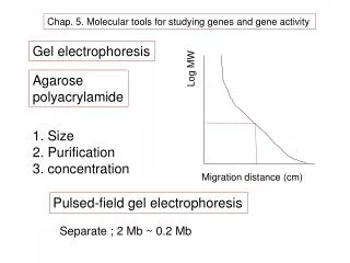
Chapter 5 Molecular Tools for Studying Genes and Gene Activity. Jay D. Hunt, Ph.D. Department of Biochemistry and Molecular Biology CSRB 4D1 568-4734 jhunt@lsuhsc.edu. Electrophoresis Agarose gel electrophoresis. Separates DNA (or RNA or Protein) fragments on the basis of charge and size

E N D
Chapter 5 Molecular Tools for Studying Genes and Gene Activity Jay D. Hunt, Ph.D. Department of Biochemistry and Molecular Biology CSRB 4D1 568-4734 jhunt@lsuhsc.edu
Electrophoresis • Agarose gel electrophoresis
Separates DNA (or RNA or Protein) fragments on the basis of charge and size • Because DNA is an acid, it looses protons in basic buffers; thus it has a negative charge that is uniform per unit length • Agarose (a polysaccharide) or other gel matrices are difficult for large DNA fragments to move through • The larger the fragment, the more difficulty it has moving through gels • By placing DNA in a gel, then applying a voltage across the gel, the negatively charged DNA will move toward the anode (positive electrode) • Large fragments lag behind while small fragments move through the gel relatively rapidly
Gel Electrophoresis - Wells Direction of DNA Travel + Large Small
Text Art Page 91 The electrophoretic mobility of a DNA fragment is inversely proportional to the log of its size.
Figure 5.2b 3 mm 10 20 30 40 50 60 70
Electrophoresis • Agarose gel electrophoresis • PFGE
+ + + + + | | | | | But, what if you want to separate larger fragments, such as entire yeast chromosomes? Pulsed-field gel electrophoresis (PFGE) can resolve fragments from 200 Kpb (0.2 Mbp) to 6000 Kbp (6 Mbp).
Figure 5.3 2.2 Mbp 0.2 Mbp
Electrophoresis • Agarose gel electrophoresis • PFGE • SDS-PAGE
Electrophoresis of proteins using SDS-polyacrylamide gel electrophoresis (SDS-PAGE) • Denaturing electrophoresis • Detergent (SDS) • Reducing agent (b-mercaptoethanol) • Heat • SDS binds to denatured proteins, making them negatively charged • Migrate through gel based on size • Molecular weight markers allow for estimation of size of polypeptide • Modifications (e.g., glycosylation) can significantly impact the apparent size of the protein
Polyacrylamide gels are composed of chains of polymerized acrylamide that are cross-linked by a bifunctional agent • N,N’-methylene-bis-acrylamide • Size of pores decrease as the ratio of bisacrylamide:acrylamide increases, reaching a minimum at ~1:20 ratio • A 1:29 ratio is most commonly used, as it is capable of resolving polypeptides that differ is size by as little as 3%
Electrophoresis of proteins using SDS-PAGE Molar ratio of bisacrylamide:acrylamide is 1:29
Electrophoresis • Agarose gel electrophoresis • PFGE • SDS-PAGE • 2-D gels
+ + Separated by isoelectric point Acidic Separated by Size Proteins stop migrating when they reach their isoelectric point (pH at which they have no net charge) Isoelectric focusing Basic | | Standard SDS polyacrylamide gel
Treat cells with a drug Treat cells with vehicle (control) Label proteins with Cy5 (blue) Label proteins with Cy3 (red) 2D-gel 2D-gel Overlay gels Analyze for differences
Electrophoresis • Agarose gel electrophoresis • PFGE • SDS-PAGE • 2-D gels • Other types of chromatography • Ion-exchange chromatography
Low Salt High Salt DEAE-Sephadex (Positively Charged) Negatively charged proteins bind to resin (the stronger the charge, the tighter the binding) Tighter bound protein elutes with higher concentrations of salt Weakest bound protein (weakest negative charge) comes off first.
Electrophoresis • Agarose gel electrophoresis • PFGE • SDS-PAGE • 2-D gels • Other types of chromatography • Ion-exchange chromatography • Gel filtration chromatography
Buffer Sephadex Smaller proteins elute with additional buffer Largest proteins come off first (void volume).
Electrophoresis • Agarose gel electrophoresis • PFGE • SDS-PAGE • 2-D gels • Other types of chromatography • Ion-exchange chromatography • Gel filtration chromatography • Autoradiography • X-ray film
X-ray Film Cassette Intensifying Screen: •b-emitters only •(3H, 14C, 35S, 32P) •-70°C or cooler X-ray Film Nitrocellulose or Nylon Membrane
Ag Ag Ag Ag Ag Ag Ag Ag Ag Ag Ag Ag 32P 32P Intensifying screen (fluoresces with b-rays)
Figure 5.9 Densitometry
Electrophoresis • Agarose gel electrophoresis • PFGE • SDS-PAGE • 2-D gels • Other types of chromatography • Ion-exchange chromatography • Gel filtration chromatography • Autoradiography • X-ray film • Phosphorimaging
X-ray film cassette Storage Phosphor plate Nitrocellulose or Nylon Membrane
Phosphorimaging is much more sensitive than X-ray film • <0.95 dpm/mm2 for 1 hr exposure to 14C • <0.15 dpm/mm2 for 1 hr exposure to 32P • Dynamic range of 5 orders of magnitude • Shorter exposure times (50-90%)
Electrophoresis • Agarose gel electrophoresis • PFGE • SDS-PAGE • 2-D gels • Other types of chromatography • Ion-exchange chromatography • Gel filtration chromatography • Autoradiography • X-ray film • Phosphorimaging • Liquid scintillation counting
Electrophoresis • Agarose gel electrophoresis • PFGE • SDS-PAGE • 2-D gels • Other types of chromatography • Ion-exchange chromatography • Gel filtration chromatography • Autoradiography • X-ray film • Phosphorimaging • Liquid scintillation counting • Non-radioactive tracers
Electrophoresis • Other types of chromatography • Autoradiography • Nucleic acid blotting • Southern blotting
Southern blot: transfer of DNA from a gel to a solid support membrane Northern blot: transfer of RNA from a gel to a solid support membrane
Electrophoresis • Other types of chromatography • Autoradiography • Nucleic acid blotting • Southern blotting • DNA fingerprinting
Electrophoresis • Other types of chromatography • Autoradiography • Nucleic acid blotting • Southern blotting • DNA fingerprinting • Northern blotting


![[II] Molecular Techniques for Studying Gene Expression](https://cdn2.slideserve.com/4213024/ii-molecular-techniques-for-studying-gene-expression-dt.jpg)