Download
1 / 24
240 likes | 375 Views
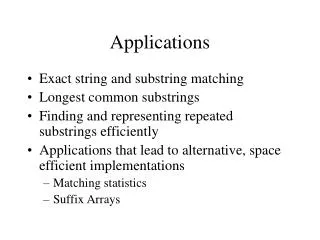
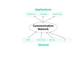
Applications. cbEGF32-33 with and without Ca 2+ c Refinement using T 1 /T 2 ratios in 4 F1- 5 F1 Peptide Binding in 1 F1- 2 F1. N. N. C. C. TB-6. cbEGF32-33. Domain Organisation of Human Fibrillin-1. TB-6. cbEGF32-33. Fibrillin is Part of the EM . Fibrillin Microfibril

E N D
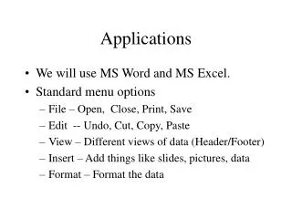
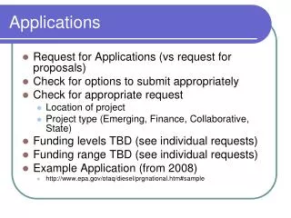
Applications • cbEGF32-33 with and without Ca2+c • Refinement using T1/T2 ratios in 4F1-5F1 • Peptide Binding in 1F1-2F1
N N C C TB-6 cbEGF32-33 Domain Organisation of Human Fibrillin-1 TB-6 cbEGF32-33
Fibrillin is Part of the EM Fibrillin Microfibril Extracellular Matrix
Model for Microfibril Organisation = putative MAB binding site
Ca2+ modulates overall structure EM Micrographs of Microfibrils with Ca2+ with EDTA
tm= 5.1 ns D||/D = 1.55 sin2(a) Ca2+ bound cbEGF32-33 is elongated
Relaxation of cbEGF32-33:with and without Ca2+ cbEGF32 cbEGF33 • local motions dominate cbEGF32 cbEGF33
N Ca2+ bound Ca2+ free N C C Removal of Ca2+ Increases Slow Motion Rex > 8 Hz Rex 4-8 Hz Rex 2-4 Hz
Model of the effect of removing Ca2+ Wobble model
10F3 1F2 1F1 Modular Structure and Binding Sites of Fibronectin Cross Linking EDA EDB Site IIICS COOH RGD s s NH 2 COOH Fibrin Cell Heparin Collagen Fibrin Gelatin S. aureus C1q Heparin
family of refined structures (without T1/T2) for each structure (i) fit T1/T2 data to obtain a c2(i) high c2(i) “bad” structures intermediate c2(i) structures low c2(i) “good” structures Use of T1/T2 Data in Analysis rank structures according to c2(i) classify structures
Ranking of Structures: 4F15F1 8 structures with high c2 8 structures with intermediate c2 8 structures with low c2 RMSD4F1= 1.04 Å RMSD5F1= 9.38 Å RMSD4F1= 1.10 Å RMSD5F1= 8.62 Å RMSD4F1= 1.07 Å RMSD5F1= 4.40 Å
Refinement with T1/T2 Potential Tjandra et al. (1997) Nat.Struct.Biol4, 443 • simulated annealing (Xplor(v3.8) or CNS (0.9)): Epot(r)=Ebonded(r)+Enonbonded(r)+Eexp(r)experimental: Eexp(r)=ENOE +Edihedral+ET1/T2 • T1/T2 potential: ET1/T2= kDANI [(T1/T2calc)-(T1/T2exp)]2
Optimisation of Parameters A) force constant: B) diffusion tensor C) SA protocol?
In vacuo In vacuo & T1/T2 4F1 5F1 Structure Refinement of 4F15F1 Inclusion of T1/T2 restraints improves definition of structure
1F1 2F1 1F1 2F1 B3 peptide B3 peptide 1F1 2F1 • AUC • ITC B3 peptide B3 forms a 1:1 Complex with 1F12F1
1F1 1F1 2F1 2F1 B3 peptide B3 Peptide Increases Anisotropy
1F1 2F1 1F1 2F1 B3 peptide B3 peptide 1F1 2F1 • AUC • ITC B3 peptide B3 forms a 1:1 Complex with 1F12F1
1F1 1F1 2F1 2F1 B3 peptide Ligand induced tethering
Ligand tethering: Structures Complex NOE,RDC,T1/T2 free Complex (NOE)
Summary • Relation ship between relaxation and dynamics • T1, T2 etc depend on J(w) at specific frequencies • motional models (diffusion) provide theoretical J(w) • fit T1,T2 to motional models via J(w) • extract correlation times and amplitudes