Download
1 / 47
470 likes | 629 Views
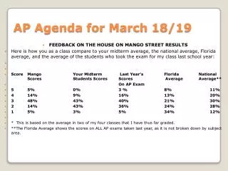
Average = 112 = B-. A = 155+ C+=100-109 A-=145-154 C=80-99 B+=135-144 C-=70-79 B=121-134 B-=110-120. Voet: Suggested Problems Ch 10: 1, 3, 4, 5, 6, 8, 10, 11, 12, Segel: Chapter 1: 53, 55 2: 1, 2, 13, 30, . Suggested problems chapter 13: 3, 4, 5, 7, 8 Chapter 14:

E N D
Average = 112 = B- A = 155+ C+=100-109 A-=145-154 C=80-99 B+=135-144 C-=70-79 B=121-134 B-=110-120
Voet: Suggested Problems Ch 10: 1, 3, 4, 5, 6, 8, 10, 11, 12, Segel: Chapter 1: 53, 55 2: 1, 2, 13, 30,
Suggested problems chapter 13: 3, 4, 5, 7, 8 Chapter 14: 3, 4, 5, 6, 12
Figure 10-29 The species and reactions permitted under the symmetry model of allosterism. Page 347
Figure 10-36 The sequential and the symmetry models of allosterism can provide equally good fits to the measured O2-dissociation curve of Hb. Page 351
Common features of enzyme active sites: 1. The active site is a 3-dimensional cleft formed from amino acids at distant sites in the sequence. 2. The active site accounts for a relatively small part of the total volume of the protein. 3. Substrates are generally bound to enzymes by non-covalent interactions. 4. The specificity of S binding depends on the arrangements of atoms in the active site.
Figure 10-35 Sequential binding of ligand in the sequential model of allosterism. Page 351
What are enzymes? • Usually proteins (exception = ribozymes) • Biological Catalyists • Facilitate reactions under physiological conditions that would require very harsh conditions in the laboratory
Enzymes biological catalysts responsible for supporting almost all of the chemical reactions that maintain organismall homeostasis. The macromolecular components of almost all enzymes are composed of protein, except for a class of RNA modifying catalysts known as ribozymes. Ribozymes are molecules of ribonucleic acid that catalyze reactions on the phosphodiester bond of other RNAs. Almost every significant life process is dependent on enzyme activity.
Functions of Catalysts • Lower the Ea for a reaction • Are regenerated • Do not affect equilibrium position
Table 13-3 Enzyme Classification According to Reaction Type. Page 470
Figure 13-1 An enzyme–substrate complex illustrating both the geometric and the physical complementarity between enzymes and substrates. Page 460
Number Classification Biochemical Properties 1. Oxidoreductases Act on many chemical groupings to add or remove hydrogen atoms. 2. Transferases Transfer functional groups between donor and acceptor molecules. Kinases are specialized transferases that regulate metabolism by transferring phosphate from ATP to other molecules. 3. Hydrolases Add water across a bond, hydrolyzing it. 4. Lyases Add water, ammonia or carbon dioxide across double bonds, or remove these elements to produce double bonds. 5. Isomerases Carry out many kinds of isomerization: L to D isomerizations, mutase reactions (shifts of chemical groups) and others. 6. Ligases Catalyze reactions in which two chemical groups are joined (or ligated) using ATP.
1.Addition or removal of water a.Hydrolases - these include esterases, carbohydrases, nucleases, deaminases, amidases, and proteases b.Hydrases such as fumarase, enolase, aconitase and carbonic anhydrase 2.Transfer of electrons a.Oxidases b.Dehydrogenases 3.Transfer of a radical a.Transglycosidases - of monosaccharides b.Transphosphorylases and phosphomutases - of a phosphate group c.Transaminases - of amino group d.Transmethylases - of a methyl group e.Transacetylases - of an acetyl group 4.Splitting or forming a C-C bond a.Desmolases 5.Changing geometry or structure of a molecule a.Isomerases 6.Joining two molecules through hydrolysis of pyrophosphate bond in ATP or other triphosphate a.Ligases
Oxidoreductases--catalyze redox reactions Usually require a coenzyme Ethanol + NAD+ Acetaldehyde + NADH + H+ Enzymes receive “common” names reflecting their function, either in the forward or reverse direction. The enzyme for this reaction is called Alcohol Dehydrogenase
Transferases-transfer functional groups Kinases transfer phosphates from ATP (or GTP) E.g Hexokinase: Glucose + ATP glc-6-P + ADP Hydrolases catalyze hydrolytic cleavages Proteases are hydrolases
Lyases catalyze group elimination to form double bonds e.g. Enolase (glycolysis) 2-Phosphoglycerate H2O + phosphoenolpyruvate Isomerases--duh, interconvert isomers e.g. phosphoglucose isomerase Glucose-6-phosphate Fructose-6-phosphate
Ligases--join to substrates together at the expense of ATP e.g. DNA Ligase Joins Okazaki fragments during DNA replication Some bacterial ligases substitute NAD+ as the energy source.
Coenzymes • Enzymes often require the participation of other small molecules to carry out a particular reaction. • These small molecules, called coenzymes, are metabolic derivatives of vitamins. • Vitamins are nutrients required in small amounts by organisms. Vitamin deficiencies usually present as metabolic disorders, e.g. scurvy
Figure 13-2 The structures and reaction of nicotinamide-adenine dinucleotide (NAD+) and nicotinamide adenine dinucleotide phosphate (NADP+). Page 461
Figure 13-4 Structures of nicotinamide and nicotinic acid. Page 464
Enzyme Activities Are Regulated at Various Levels • Transcription • Processing • Translation • Post-translational modification • Transient modification (e.g. phosphorylation) • Allosteric Effectors
Figure 13-5 The rate of the reaction catalyzed by ATCase as a function of aspartate concentration. Page 465
Figure 13-6 Schematic representation of the pyrimidine biosynthesis pathway. Page 466
Figure 13-7a X-Ray structure of ATCase. (a) (left) T-state ATCase along the protein’s molecular threefold axis of symmetry; (right) R-state ATCase along the protein’s molecular threefold axis of symmetry. Page 467
Figure 13-7b X-Ray structure of ATCase. (b) (left) T-state ATCase along the protein’s molecular twofold axis of symmetry; (right) R-state ATCase along the protein’s molecular twofold axis of symmetry. Page 467
Figure 13-8 Comparison of the polypeptide backbones of the ATCase catalytic subunit in the T state (orange) and the R state (blue). Page 468
Figure 13-9 Schematic diagram indicating the tertiary and quaternary conformational changes in two vertically interacting catalytic ATCase subunits. Page 469
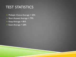
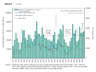
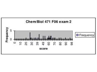
Average= 72.8 Standard Dev=11.7
A 170 + B137-166 C 110-136