Download

1 / 33
350 likes | 565 Views
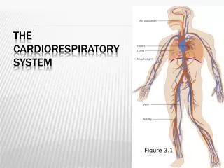
Cardiorespiratory system. Cardiovascular System Respiratory System. Transportation of nutrients, gases, and waste Protection from infection and blood loss Maintenance of body temperature Supply blood with oxygen Removal of carbon dioxide. Cardiovascular system.

E N D
Cardiovascular SystemRespiratory System • Transportation of nutrients, gases, and waste • Protection from infection and blood loss • Maintenance of body temperature • Supply blood with oxygen • Removal of carbon dioxide
Cardiovascular system • BLOOD VESSELS “the roads, railway tracks, and airways of the body” • Arteries • Veins • Capillaries BLOOD VESSELS “the roads, railway tracks, and airways of the body” Arteries “freeway” – carry blood AWAY from the heart Veins – carry blood TOWARDS the heart Capillaries “side streets”– tiny, ‘hair-like’ blood vessels where gas exchange occurs
Cardiovascular system • Heart – pump of the body • About the size of your fist • Your heart is a muscle (remember involuntary cardiac muscle!) that contracts and relaxes automatically approximately 70 beats per minute at rest • The heart is divided into a right and left half – each are pumps and ensure there’s no mixing between oxygenated blood and deoxygenated blood • Each half of the heart is in charge of pumping blood through different blood vessel circuits
THE HEArt • 4 chambers of the heart • Atria – right/left • Ventricles – right/left • Valves – ensure blood flows in a one • direction • tricuspid, pulmonary, mitral (bicuspid), aortic • Veins – carry blood TOWARDS the heart • Superior Vena Cava • Inferior Vena Cava • Pulmonary Vein* • Arteries – carry blood AWAY from the heart • Pulmonary Artery* • Aorta • Septum – separates the left and right ventricles septum
Blood flow through the heart • Oxygenated blood enters the left atrium through the pulmonary veins* • The left atrium pumps the blood through the mitral valve into the left ventricle • The left ventricle contracts and pumps the blood through the aortic valve towards the body systems where the oxygen is delivered through the capillaries • The deoxygenated blood is transported back towards the heart through the veins. The superior and inferior vena cava both transport deoxygenated blood to the right atrium • The right atrium contracts and pumps the blood through the tricuspid valve • The right ventricle pumps the blood through the pulmonary valve to the pulmonary artery* which delivers the deoxygenated blood to the lungs where it become oxygenated blood through gas exchange in the capillaries • The pulmonary vein then transports the oxygenated blood to the left atrium where the cycle is continued (about 70 times per minute!) • Blood flow videoBlood flow video 2 *????????
What makes the ‘lub-dub’ noise? • Lub • Atria contract to fill ventricles • Ventricles are full • Tricuspid (T) and mitral (M) valves snap shut to prevent backflow • Pulmonary (P) and aortic (A) valves are open • Dub • Ventricles contract • Blood flows through pulmonary (P) and aortic (A) valves • Pulmonary (P) and aortic (A) valve snap shut to prevent backflow • Tricuspid (T) and mitral (M) valves are open Lub – T+M snap shut, P+A open Dub – P+A snap shut, T+M open
NOTEBOOk heart Systole and Diastole
ACTIVITY TIME • Turn to page 36 in your PINK notebook • LABEL THE HEART • COLOUR THE OXYGENTATED HALF IN RED AND THE DEOXYGENTATED HALF IN BLUE • 2. Time-permitting: in groups of 3, create a song, skit, or comparison to explain the blood flow through the heart.
When you breathe in (inhale) air is carried through your air passages and into your lungs, expanding them like a blown-up balloon. The expansion of your lungs would not be possible without respiratory muscles, such as pectoralis major and minor and the diaphragm. Pectoralis major and minor (chest muscles) lift your ribs up and out to make room for the lungs to expand
The diaphragm is the strong muscle that separates the chest cavity from the abdominal cavity. When your diaphragm moves down, your lungs expand and suck in air. When you exhale, your diaphragm rises to squeeze carbon dioxide out of your lungs
Pathway of Respiration • Nasal/Oral cavity Pharynx Larynx Trachea Bronchi (left and right lung) Bronchioles Alveoli
***Around each alveolus there is a network of blood vessels called capillaries. Oxygen from inhaled air passes into the capillaries, while carbon dioxide from blood that has already circulated around the body is passed back into the lungs to be exhaled.
Smoking What do you think about when you hear the word SMOKING??????
Smoking is hazardous to all of the systems in your body, especially your respiratory system. When you smoke a cigarette, you inhale the smoke into your lungs. This means that your lungs are directly exposed to the 4,000 toxic substances in cigarette smoke. These substances can impair your lungs' ability to function and interfere with the mechanisms that protect your lungs against disease.
Closer look at Respirations: Respiratory Rate (RR) • Each respiration involves one inhalation (chest rises) and one exhalation (chest falls) • Normally quiet, effortless and regular (both sides of the chest rise and fall equally) • Factors that affect respiration rate: age, weather, exercise, pregnancy, menstrual cycle, emotions and stress • If the chest barely moves, the breathing is called shallow • If the chest rises and falls significantly with every breath, this is known as deep breathing • Count how many respirations the client has in 30 seconds and then multiply it by 2 (table 40-3 (pg. 667) – Normal Respiratory Rates by Age) • Copy and practice procedure on pg. 668