Download
1 / 1
10 likes | 106 Views
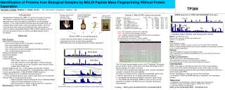
Identification of Proteins from Biological Samples by MALDI Peptide Mass Fingerprinting Without Protein Separation. TP389. Kenneth C. Parker , Stephen J. Hattan, Jie Du. VIC Instruments Corporation, Sudbury , MA. MSMS spectrum of YPIE h GIITNWDDMEK from actin. Home-brewed beer

E N D
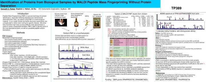
Identification of Proteins from Biological Samples by MALDI Peptide Mass Fingerprinting Without Protein Separation TP389 Kenneth C. Parker, Stephen J. Hattan, Jie Du.VIC Instruments Corporation, Sudbury , MA MSMS spectrum of YPIEhGIITNWDDMEK from actin Home-brewed beer (from daughter’s boyfriend) Typical LC-MALDI PMF results from chicken Grocery store food (chicken thigh and breast meat) Introduction Generic Art (oil / tempera) h saliva Peptide Mass Fingerprinting (PMF) is a proven technique for protein identification, especially following separation by 2-dimensional electrophoresis. Here, we explore using PMF on crude biological material. In our model chicken biomarker discovery experiments, we compare dark to white meat, looking for specific masses. We show here that when high mass accuracy MALDI mass spectrometry is used on certain unseparated samples, PMF is able to identify some of the most abundant proteins, as well as candidate peptide isoform biomarkers. P or R L M E W Y W y1 b2 or ME hG PI hGI a5 EhG b6 b4 b5 #Obs -the # of peptides matched, after subtraction. #Obs_I -the initial number of peptides matched %CM -related to coverage, but weighted toward Arg-containg peptides. %IM - percentage intensity matched. All these proteins confirmed by LC-MALDI MSMS. 27 replicate digests of chicken meat (13 white, 14 dark) h indicates methyl histidine, with strong proton affinity Methods Perform PMF on un-purified protein • Saliva-> (by PMF only so far.) • Amylase (22% intensity), cystatin, IgA (supernatant) • keratin 4, 13, 6A (pellet) . • Beer-> • yeast glycolytic enzymes • barley ~protease inhibitors (at least 3 isoforms each) by both PMF and MS-MS • PMF IDs marginal compared to the other systems, probably due to incomplete database issues, or protein degradation • Painting -> • egg white (ovalbumin, lysozyme) from pigeon or duck. • work supplied by and corroborated independently of us by Dan Kirby and Katherine Phillips, Harvard U. • Reagent contamination-> • identifies EfTu, porin from Burkholderia species. • Accomplished by starting from SwissProt Bacteria database, • then working through Trembl using smaller phylogenetic categories. Chicken dark and white meat is a model system for characterizing human muscle biopsy samples. What differences can be observed without protein separation? • PMF Analysis • Get protein-containing sample • -from chicken breast (white) or leg (dark); homogenize • - from home-brewed beer sediment • - from saliva (supernatant or pellet) • -from oil-painting in museum (prepared by Dan Kirby, Harvard U.) • -from contaminated lab stock solution • Reduce and alkylate in SDS • Acetone Precipitate • Trypsin Digest • Collect MS Spectra • Use ‘combo’ machine-> 20,000 resolution • Use ‘elite’ machine, 14.8 m flight path-> 45,000 resolution • 1 or 2 kHz laser (up to 4 khZ possible, starts failing at 5kHz) • Collect 4000-10000 shots • Prepare Peak list with 100 -~1000 masses, density filter if desired. • Perform PMF Vs. combined SwissProt / Trembl database, keyed to taxon • Confirm as needed with LC MALDI MSMS (MSMS machine). • Prepare Peak List from LC-MALDI composite spectrum • or from LC-MALDI peak list (also used to select precursors for MS-MS) • Perform PMF on these peak lists as well. • Nearly equivalent results obtained as from unseparated digest • Useful for testing calibration across LC run. • Best results with LC-MALDI peak list from 45 K resolution! White Meat Dark Meat • Conclusions: • PMF is successful in identifying proteins from many crude samples. • At top level, MYH isoforms distinguish chicken white meat from dark. • Annotating database sequences with quantitative modifications (like actin h shown above) aids PMF. • Protein degradation is often less serious than expected. • Confirmation of PMF by MSMS requires LC separation (of homologous sample). • The 100 most intense masses found in the 27 replicates. 26 masses were enriched in dark or white meat, and mostly matched to isoform-specific peptides from MYH of chicken. • The most intense peptide was mapped to actin, and was expressed ~equally, as were all other actin peptides. • Sc refers to the Mascot Score, searched using MSMS spectra from LC-MALDI experiments Vs. the NCBI vertebrate database. • p -the peptide was identified by MSMS in at least 2 variant forms. • m- the peptide was identified by MSMS in only one form, but the sequence is variable in the MYH isoforms that were identified by other peptides. References: 1.) Parker KC. Scoring Methods in MALDI Peptide Mass Fingerprinting: ChemScore and the ChemApplex Program. JASMS 2002;13:22-39. 2.) See Poster WP651 for more details. 1360 1410 Abbreviations: myosin heavy chain -> MYH Several MYH isoforms are evident that differ between white and dark meat. Funding: SBIR grants 5R44RR025705, 5R44GM079832; SBIR grants 5R44GM079833, 1R43RR030734