Download
1 / 46
500 likes | 1.04k Views
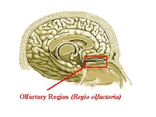
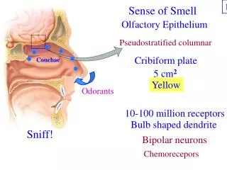
Sense of Smell. 1. Odorants . Sniff!. Olfactory Epithelium. Pseudostratified columnar. Cribiform plate. Conchae . 5 cm 2. Yellow. 10-100 million receptors. Bulb shaped dendrite. Bipolar neurons. Chemorecepors . Microanatomy of Olfactory Epithelium. 2. Olfactory tract.

E N D
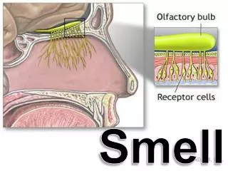
Sense of Smell 1 Odorants Sniff! Olfactory Epithelium Pseudostratified columnar Cribiform plate Conchae 5 cm2 Yellow 10-100 million receptors Bulb shaped dendrite Bipolar neurons Chemorecepors
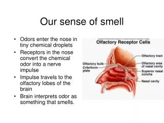
Microanatomy of Olfactory Epithelium 2 Olfactory tract Olfactory Bulb Olfactory bulb neurons Cribiform plate Olfactory nerve (I) Olfactory gland Basal stem cell Olfactory receptor Support cells Dendrite Mucus Olfactory hairs Odorants enter 2nd order 1st order 60 days
Olfactory Receptor Activation 3 Unique receptors Receptors Transduced Adenylate cyclase Receptor potential cAMP Action potential Na+ channels Ca+2 channels Olfactory bulb neuron Adaptation Receptor potential 1000 genes Several odors Odorants G-protein
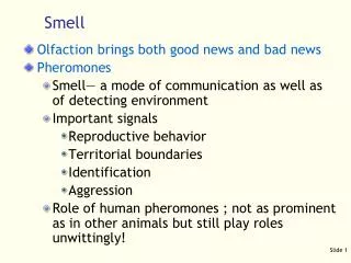
Olfactory Pathway 4 olfactory tract “fight or flight” Parasympathetic Olfactory bulb neurons (2nd order) 2 main destinations: Olfactory cortex Path 1: Thalamus (3rd order) #11 frontal lobe Identification Path 2: Hypothalamus Amygdala Other Limbic Emotional response Smells of danger Appetizing odors Protective responses Sneezing!
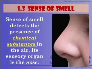
Sense of Taste 5 Chemical sense 5 primary tastes: Sour (Acids) Salty (Metal ions) Bitter (Alkaloids) Sweet (Sugars) Umami (Glu, Asp) All other flavors: Combinations of 5 tastants Olfactory sensations Tactile Pain
Taste Bud 6 Epiglottis Palatine tonsil Lingual tonsil Vallate Papilla Foliate papilla Filiform papilla Fungiform papilla Papillae 8 - 12 100-300 taste buds Pointed, thread-like Taste buds on top mushroom Tactile
Taste Buds 7 Vallate papilla Filiform papilla Fungiform papilla Taste pore Gustatory hairs Stratified squamous Support cells Gustatory receptor cells Basal cells 1st order dendrites Chemoreceptors
Fungiform papillae http://www.iamfightingcancer.com/tongue%20taste%20buds.jpg http://anatomy.iupui.edu/courses/histo_D502/D502f04/Labs.f04/digestiv%20I%20lab/s41.20x.ug.jpg 8
Taste Receptor Activation 9 Receptors on gustatory hairs Transduction Receptor potential Neurotransmitter release Receptors on 1st order neurons Na+ channels Depolarization Opens Ca+2 channels Synaptic vesicles fuse Tastants Salty food
Gustatory Pathway 9 ANS Reflexes: Thalamus (3rd order) Gustatory cortex 1st order neurons Anterior 2/3 Posterior 1/3 Epiglottis & pharynx Facial Glossopharyngeal Vagus Medulla (2nd order) Saliva Gastric juices Gagging Limbic system Appreciate! Taste aversion Conscious perception
Eye Accessory Structures 11 Lysozyme Lacrimal gland Lacrimal caruncle Lacrimal sac Meibomian glands Lacrimal fossa Sebaceous ciliary glands Bulbar conjunctiva Bloodshot Mucus Lacrimal apparatus: Lacrimal fluid Eccrine sweat glands Sebaceous glands Palpebrae Tarsal plates Oily (Chalazion) (Sty) Palpebral conjunctiva
Fibrous Tunic 11 Cornea Sclera Extrinsic muscle (transparent) Avascular Protect the Iris Tough, fibrous CT Focus light on retina Transplant Lens Shape Protects (III, IV, VI)
Vascular Tunic Ciliary body 12 Zonular fibers Iris Ciliary processes Pupil Ciliary muscle Ora serrata Choroid Uvea Suspend the lens Regulate light Aqueous humor Alters lens shape Lens Anterior margin Retina Nutrients to all tunics Absorbs light
Iris functions 13 Radials contract Circulars contract 2 sets smooth muscle: Control size of pupil Bright light, close vision Dim light, distant vision Normal Dilate Constricts Sympathetic Parasympathetic
Lens 14 Biconvex disc Transparent Flexible Avascular Simple cuboidal epithelium Differentiate into lens fibers: Anucleate Crystallins Function: Active focusing
Image Formation for close vision 15 Zonula goes slack Brings divergent rays together Curvature Accommodation - Increases refractory power of lens Ciliary muscles contract Lens bulges round Pupil constriction Circular smooth muscle contracts Narrows diameter Prevents entry of divergent light Convergence - Medial rotation of eyes Somatic motor - III Eyestrain
Sensory tunic – 2 layers Pigmented epithelium Visual pathway Retina Optic disc Central fovea Absorb light Neural layer Store vitamin A Phagocytes Rods Photoreceptors Macula (Optic nerve exits) (Cones only) 16
Neural Layer Pigmented epithelium Cones Photoreceptor layer Rods Receptor potential Bipolar cell layer Visual acuity Ganglion cell layer Optic nerve Melanin Color Night Transduce light Macula lutea Visual axis Central fovea Integration Transmit Light Optic disc 17
Photoreceptor - Rod 18 Pigmented epithelium Phagocytized daily Outer segment Inner segment Synaptic terminal New discs Membrane discs Intense light damages 100 rods to 1 bipolar Out put is gray tones Produce photopigments Absorb all wavelengths Vitamin A Very sensitive – night vision Photopigment - Rhodopsin
Photoreceptor - 3 Cones 19 Pigmented epithelium Outer segment Renewal at night Low sensitivity Bright light stimulates 3 photo-pigments = color Pleated & continuous 3 primary colors Red – 560 nm Blue – 420 nm Green – 530 nm 1 cone = 1 bipolar High resolution
Eye Interior 20 Cornea Anterior cavity Posterior cavity Aqueous humor Continuous Nourish lens & cornea Glaucoma Lens (Lifetime) Vitreous humor Transmits light Intraocular pressure Holds retina against choroid Prevents detachment Even surface for image formation
Visible Light 21 Wavelength 400 – 700 nm 1st to 2nd medium Refracted Central fovea Photons Vibrating packet of pure energy Bright wiggle Ultraviolet Infrared Colored objects: absorb some wavelengths reflect others to cones Speed of light changes Light bends Light strikes the cornea & lens it refracts 3X Focuses the light
Pathway of Light 22 Cornea – aqueous humor – lens – vitreous humor Thru entire thickness of retina to excite photoreceptors 3 refractions: Cornea 75% Entering & leaving the lens 25%
Photoreceptor Status at Rest 23 Na+ enters = Dark current Partially depolarizes - 40 mV Voltage-gated Ca+2 Continual release glutamate Inhibitory PSP - Bipolar neurons DARK! cGMP binds Na+ channels Photoreceptors & bipolar signal by graded potentials
Stimulation of Rhodopsin 24 Retinal changes shape Releases opsin G-protein (transducin) Enzymes degrade cGMP Na+ channels close K+ = photoreceptor hyperpolarizes Photopigments contain 2 compounds: Retinal - Vitamin A Light absorbing component Opsin - Protein Retinal + 1 photon Blockade removed
Visual Pathway 25 Ganglion cells = action potential Optic chiasma Left thalamus Right thalamus Bipolar depolarizes Optic nerve Visual cortex Superior colliculi Hypothalamus Form, color movement Pupilary reflexes Circadian Rhythms Extrinsic reflexes Visual association Identification Spatial location
26 Few nutrients to lens fibers ReStore multifocal lens Cataracts Hardening, thickening of lens >Age Diabetes mellitus >Smoking Crystallins clump >Sunlight Artificial lens Contacts, glasses
Glaucoma 28 Retina, optic nerve Canal of Schlemm Halos, blurred vision Congenital weakness of extrinsic Dangerous intraocular pressure Failure to drain aqueous humor Slow Eye drops, laser surgery Strabismus Cross-eyed Vision from one eye 2nd functionally blind Exercises, patching, surgery
Anatomy of the Outer Ear 29 Pinna External auditory meatus Tympanic membrane Funnel sound waves Ceruminous gland Wax Vibrate
Middle Ear 30 Oval window Malleus Incus Stapes Auditory tube Mucosa Filled with air Waves in perilymph Ossicles: Membrane pulses Amplify vibrations from tympanic membrane Equalizes pressure Mucosa = middle ear + pharynx
Inner Ear Semicircular canals Vestibule Cochlea Bony labyrinth in temporal bone Perilymph Membranous labyrinth Endolymph Vestibular Apparatus: 2 components Dynamic equilibrium Static equilibrium Hearing 31
Vestibular Apparatus 32 Anterior Posterior Lateral Semicircular canals Utricle Maculae 3 Ampulla Saccule Vestibule Receptor organs for equilibrium Static Head position Crista Dynamic – 3D Rotary head movements Oval window
Macula - Static Otoliths – Ca2CO3 Inertia 33 Hair bundle Glycoproteins 1st order neuron Hair cells Support cell Epithelium Stereocilia Receptor Nurture receptors Otolithic membrane VIII - Vestibular branch
Macula Monitor position of head in space 34 Otoliths move with gravity Otoliths Otolithic membrane Hair bundles Receptor potential Hair cells Bent Forward Straight-line changes Saccule & Utricle Bend Straight up 1st order Upright
Crista Stereocilia Crista Hair cells Support cells 1st order neuron Dynamic Rotarymovements Change in velocity 3 Ampulla– 1 for each plane Endolymph Cupula 35
Activation of Crista 36 Head moves Semicircular canals & hair cells move too Endolymph lags behind Drags the Cupula backward Bends hair bundles Receptor potential
Cochlea 37 Cochlear duct Basilar membrane Vestibular membrane Scala vestibuli Scala tympani Temporal bone Cross section Endolymph Perilymph Perilymph Spiral, cone shaped, bony chamber
Cochlea 38 Enters Scala tympani Stapes Cochlear duct Helicotrema Round window Scala vestibuli Return to vestibule Waves in perilymph
Cochlea 39 Cochlear duct Tectorial membrane Organ of Corti Spiral ganglia Basilar membrane Scala vestibuli Perilymph Endolymph 1st order Scala tympani Perilymph
Organ of Corti 40 Hair bundles Hair Cells Support cells 1st order Basilar membrane Vibrate Scala tympani epithelium Bend Release neurotransmitter Tectorial membrane Receptor cells Waves in Perilymph
Sound waves 41 External acoustic meatus Outer ear Tympanic membrane Malleus Incus Middle ear Stapes Oval window Perilymph of scala vestibuli Perilymph of scala tympani Inner ear Basilar membrane vibrates Moves hair cells Receptor potential
Auditory Pathway 42 Neurotransmitter Hair receptor cells (VIII cochlear branch) 1st order (spiral ganglia) Midbrain Medulla 2nd order Inferior colliculi Auditory reflexes Thalamus Primary auditory cortex Pitch & rhythm
Pitch Frequency of a sound vibration 43 Faster the frequency = higher the pitch Faster frequency = higher energy 1 Hz = 1 cycle per second 1 second 1 2 3 4 1 second Low pitch = low frequency 1 2 3 4 5 6 7 8 1 second High pitch = high frequency Loud Quiet 20 Hz
Cochlea uncoiled 44 Scala vestibuli Scala tympani Stapes pushes oval window 20 Hz Width of basilar membrane Long & floppy Thin & stiff Low frequency Very high frequency Location of activated hair cells specific cortical neurons
Deafness - 45 Any hearing loss Sensorineural - Neural structures Cochlear hair cells Auditory cortex Loss of hair cells = Noise Conduction - Prevents mechanical transfer Perilymph Earwax Perforated eardrum Otosclerosis