Download
1 / 28
590 likes | 1.63k Views
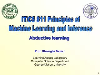
ECG & Machine Principles. ECG ELEMENTS. +. +. =. ELECTROCARDIOGRAPHER. ECG PAPER. ECG PAPER. ECG LEADS. ECG LEADS. ECG Vectors & Deflections. Frontal plane Horizontal Plane. CARDIAC Axis. Find Perpendicular. This patient’s axis is deviated to the LEFT. (– 30°) .

E N D
ECGELEMENTS + + =
ECG Vectors & Deflections • Frontal plane • Horizontal Plane
CARDIAC Axis Find Perpendicular Thispatient’s axis isdeviatedtothe LEFT. (– 30°) Find Most Isoelectric Is it positive or negative? Because aVL is POSITIVE (upward deflection on the ECG), the axis is approaching aVL, so it’s – 30°. If it was NEGATIVE on the ECG, the axis would be at the tail of the arrow, so 150°.
WHAT WE NEED TO KNOW… • PAPER DEFINITIONS • DOMINANT WAVES • AMPLITUDE • DURATION • WAVE-WAVE RELATION • WAVE-LEAD RELATION
AHA CONSIDERATIONSPatient & Machine Dependent • Lead axis & Heart Vector projectionon lead. • Lead as a vector (direction and length). Unipolar? • Strengthorsignalmagnitude variables. • Impedances. • Artifact, Filtering & Frequencycutoffs. • Movement, Position, Rib direction, Imaginary lines, Amputations, Female breasts, Implants and Obesity. • PMH. CC. CI. • Computer Interpretation.
AHA CONSIDERATIONSOperator Dependent • Skin cleaning. Retraining. • 12 Electrode TYPICAL placement. 3 lead? • Limb leads placement. • Precordial Placement of leads. • V4 – V6 horizontal plane. • V5 between V4 &V6. • Common placement errors. High, low, curved, switched. • Reproducibility? • Labeling.
ECGUtility • Arrhythmias. • Acute Coronary Syndromes (ACS). • Conduction Disturbances. • Hydro – Electrolytic (HE) Abnormalities. • Electrical & Structural Abnormalities. Also: • Monitoring Anti-ArrhythmicTreatment. • Non Cardiological Pre-Op Assesment. • Screening High Risk Activities. (Work/ sports)
ARRHYTHMIAClassifications • ABNORMALITY • Conduction • AV Blocks • Bundle Blocks • Origin • Supraventricular • Extrasystoles • Escape Beats • Junction • Ventricular • Extrasystoles • Escape Beats • HEART RATE • Tachyarrhythmias • Regular • Irregular • Bradyarrhythmias • Regular • Irregular
ARRHYTHMIAS TACHYCARDIAS • ST (r) • A. FIB. (i) • A. FLU. (r) • WPW • TSVP (r) • JT (r) • MFAT (i) • TV MONOM. (r) • TV POLIM. (i) • TDP (i) • VF (i) BRADYCARDIAS • SB • AV BLOCKS • 1° • 2° MI • 2° MII • 3° • HBB BLOCKS • TC Pacing. CA RHYTHMS • VENTRICULAR FIBRILATION • PULSELESS VENTRICULAR TACHYCARDIA • PULSELESS ELECTRICAL ACTIVITY • ASYSTOLE
ARRHYTHMIAInterpretation Method • Identify what’s normal = RHYTHM & AXIS • Establish Heart Rate (HR) = FREQUENCY • Determine Regularity = PATTERN • QRS Width = ORIGIN. • P Morphology = ATRIAL ACTIVITY • P/ QRS Ratio = AV CONDUCTION • ST & T = VENT. REPOLARIZATION
RHYTHM • Definition of Rhythm. • Sinus Rhythm • Variations of Rhythm. • Alterations of Rhythm • Long L-II
ORIGIN QRS COMPLEX • Wide VENTRICULAR • Narrow SUPRAVENTRICULAR
AURICULAR Activity:P WAVE • P WAVE - Anterograde - Retrograde
AV Conduction: PR INTERVAL Second – Degree AV Block (2 types)
ECGQT INTERVAL Useful in Tachyarrhythmias like TORSADE DE POINTS (a type of Ventricular Polimorphic Tachycardia) Torsade de Points
ECGST SEGMENT & T WAVE More useful in Acute Coronary Syndromes & HE disturbances.