Download

1 / 33
380 likes | 778 Views
DEVELOPMENT OF VERTEBRAL COLUMN & SPINAL CORD. Prof. Ahmed Fathalla Ibrahim Professor of Anatomy College of Medicine King Saud University E-mail: ahmedfathala@hotmail.com. OBJECTIVES. At the end of the lecture, students should be able to:

E N D
DEVELOPMENT OF VERTEBRAL COLUMN & SPINAL CORD Prof. Ahmed Fathalla Ibrahim Professor of Anatomy College of Medicine King Saud University E-mail: ahmedfathala@hotmail.com
OBJECTIVES At the end of the lecture, students should be able to: • List the layers of the spinal cord and its contents. • List subdivisions of mantle & marginal zones. • List meningeal layers and describe positional changes of spinal cord. • Describe development of vertebral column from sclerotomic portion of paraxial mesoderm. • Describe chondrification& ossification stages in vertebral development. • Describe spina bifida and its types.
Embryo Amniotic cavity Embryo Yolk sac
DEVELOPMENT OF NEURAL TUBE • Ectodermal cells dorsal to • notochord thickens to form • the neural plate. • A longitudinal groove develops • in the neural plate (neural groove). • The margins of the neural plate • (neural folds) approach to each • other and fuse to form the • neural tube.
DEVELOPMENT OF SPINAL CORD • The spinal cord develops from the caudal 2/3 of the neural tube
DEVELOPMENT OF SPINAL CORD The cells of neural tube form: • An inner ventricular zone of undifferentiated cells • A middle mantle zoneof cell bodies of neurons (future grey matter) • An outer marginal zone of nerve fibers or axons of neurons (future white matter)
MANTLE LAYER OF SPINAL CORD • Neurons of mantle layer (future grey matter) differentiate into: • A dorsal alar plate (future dorsal horn): containing sensory neurons • A ventral basal plate (future ventral horn): containing motor neurons • The 2 areas are separated by a longitudinal groove (sulcuslimitans).
MANTLE LAYER OF SPINAL CORD Dorsal median septum Proliferation and bulging of both alar & basal plates cause: • Formation of longitudinal dorsal & ventral median septa • Narrowing of the lumen to form a small central canal Central canal Ventral median septum
MARGINAL LAYER OF SPINAL CORD • Marginal layer increases in size due to addition of ascending, descending & intersegmental nerve fibers. • Myelinationof nerve fibers starts at 4th month & continues during the 1st postnatal period. Motor fibers myelinate before sensory fibers. • Marginal layer (future white matter) is divided into: dorsal, lateral and ventral funiculus (white column) Dorsal funiculus Lateral funiculus Ventral funiculus
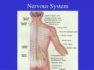
MENINGES • They are 3 membranes covering the neural tube: • Outer thick duramatter: mesodermalin origin • Middle arachnoidmatter: ectodermal in origin • Inner thin piamatter: ectodermalin origin • A cavity appears between arachnoid & pia(subarachnoid space) & becomes filled with cerebrospinal fluid.
POSITIONAL CHANGES OF SPINAL CORD • Initially, the spinal cord occupies the whole length of the vertebral canal. • As a result a faster growth of vertebral column, the caudal end of spinal cord (conusmedullaris)shift gradually to a higher level. 8 weeks: spinal cord at end of vertebral column 24 weeks: spinal cord at level of S1 Birth: spinal cord at L3 Adult: spinal cord at L1-L2
Notochord • stimulates : • Neural tube • formation • Vertebral column formation Neural tube
INTRAEMBRYONIC MESODERM • Proliferates between Ectoderm & Endoderm EXCEPT in the central axis of embryo where NOTOCHORD is found. • Differentiates into 3 parts: • Paraxial mesoderm • Intermediate mesoderm • Lateral mesoderm • Paraxial mesoderm divides into units (somites). • Each somite divides into 3 parts: • Sclerotome • Myotome • Dermatome
DEVELOPMENT OF VERTEBRA Notochord Neural tube Sclerotome Sclerotome 1 1 2 2 3 3 1- Sclerotome around neural tube: forms vertebral (neural) arch 2- Sclerotome around notochord: forms body of vertebra 3-Sclerotome in body wall near to neural tube & notochord : forms costal process (gives ribs in thoracic region)
1- Loosely arranged cells 2- densely packed cells
FORMATION OF BODY OF VERTEBRA • At 4th week, each sclerotome is formed of: • A cranial part of loosely arranged cells • A caudal part of densely packed cells • The caudal part of each sclerotome fuses with the cranial part of succeeding sclerotome to form the centrum (body primordium) • Each centrum develops from 2 adjacent sclerotomes.
FATE OF NOTOCHORD • In the region of the bodies of vertebrae: It degenerates . • Between bodies of vertebrae: It forms the intervertebral discs (nucleus pulposus). N.B.: Annulus fibrosus part of the intervertebral discs are formed by the mesoderm surrounding the notochord.
VERTEBRAL DEVELOPMENT 3 appear at end of 8th week appear at 6th week Fusion occurs at 3-5 years 5 appear at puberty Fusion occurs at 4-6 years All centers unite around 25 years
CURVATURES OF VERTEBRAL COLUMN • Primary curves (thoracic & pelvic or sacral): develop prenatally • Secondary curves: develop postnatally • Cervical:as a result of lifting the head • Lumbar: as a result of walking
SPINA BIFIDA • Cause: Failure of fusion of the halves of vertebral arches • Incidence: 0.04-0.15% • Sex: more frequent in females • Types: • Spina bifida occulta(20%) • Spin bifida cystica(80%)
Spina bifida occulta Spina bifida with meningocoele Spina bifida with meningomyelocoele Spina bifida with myeloschisis
SPINA BIFIDA OCCULTA • The closed type • Only one vertebra is affected • No clinical symptoms • Skin overlying it is intact • Sometimes covered by a tuft of hair
SPINA BIFIDA CYSTICA With meningomyelocoele With myeloschisis
SPINA BIFIDA CYSTICA • The open type • Neurological symptoms are present • Subdivided into: • Spina bifida with meningocoele: protrusion of sac containing meninges & cerebrospinal fluid • Spina bifida with meningomyelocoele: protrusion of sac containing meninges with spinal cord and/or nerve roots • Spina bifida with myeloschisis: spinal cord is open due to failure of fusion of neural folds
SUMMARY OF DEVELOPMENT OF SPINAL CORD • The spinal cord develops from the caudal 2/3 of the ectodermal neural tube. • Layers of spinal cord are (from inside outward): ventricular,mantle (future grey matter) and marginal (future white matter). • Mantle layer differentiates into dorsal alar plate (with sensory neurons) & ventral basal plate (with motor neurons) separated by sulcuslimitans. • Marginal layer is divided into dorsal, lateral & ventral funiculus.
SUMMARY OF DEVELOPMENT OF SPINAL CORD • Myelinationof nerve fibers starts at 4th month & continues during the 1st postnatal period. Motor fibers myelinate before sensory fibers. • Meningesare 3 membranous sac covering the neural tube (from outside inward): dura (mesodermal in origin), arachnoid and pia (both are ectodermal in origin). • A cavity between arachnoid & pia matters (subarachnoid space) contains cerebrospinal fluid. • During development the end of spinal cord shifts its position: at 24 weeks (level of S1), at birth (level of L3), adult position (level of L1-L2).
SUMMARY OF DEVELOPMENT OF VERTEBRAL COLUMN • Vertebral column develops from sclerotomic portion of paraxial mesoderm. • Sclerotomearound neural tube forms vertebral (neural) arch. • Sclerotome around notochord forms body of vertebra. Each body develops from 2 adjacent sclerotomes. • Notochord forms nucleus pulposus portion of the intervertebral discs. • Chondrification centers appear at 6th week. • Three primary ossification centers appear at 8th week.
SUMMARY OF DEVELOPMENT OF VERTEBRAL COLUMN • Fusion between halves of neural arch occurs at 3-5 years, between neural arch & body at 4-6 years. • Five secondary ossification centers appear at puberty and fuse around 25 years. • Spina bifida is due to failure of fusion of the halves of the neural (vertebral) arch. It may be occulta(20%, closed type, no symptoms) or cystica(80%, open type, with symptoms).
QUESTION 1 • Which one of the following regions of spinal cord contains cell bodies of sensory neurons? • Alar plate • Ventricular zone • Basal plate • Dorsal funiculus
QUESTION 2 • At which one of the following periods of life fusion between vertebral arch & body of vertebra occurs? • 8th week • Puberty • 3-5 years • Around 25 years
QUESTION 3 • Regarding spina bifidawhich one of the following statements is correct? • The closed type is more frequent than the open type. • The closed type presents with clinical symptoms. • Spina bifida is due to failure of fusion between the halves of vertebral arch. • In cases of spina bifida with meningocoele, the spinal cord is open.