Download
1 / 67
700 likes | 1.01k Views
Diseases of the Biliary Tract. Victor Politi, M.D., FACP, Medical Director, SVCMC, School of Allied Health Professions, Physician Assistant Program. Cholelithiasis (Gallstones). Cholelithiasis (Gallstones).

E N D
Diseases of the Biliary Tract Victor Politi, M.D., FACP, Medical Director, SVCMC, School of Allied Health Professions, Physician Assistant Program
Cholelithiasis (Gallstones) • Gallstone disease, or cholelithiasis, is one of the most common surgical problems worldwide. • Gallstones are abnormal, inorganic masses formed in the gallbladder and, less commonly, in the common bile or hepatic ducts
Although gallstones can form anywhere in the biliary tree, the most common point of origin is within the gallbladder. • Three types of gallstones exist: • pure cholesterol • pure pigment • mixed
Gallstones are classified according to their predominant chemical composition as either: • cholesterol • calcium bilirubinate stones • < 20% of stone type in Europe & US • 30-40% of stones in Japan
Three compounds comprise 80-95% of the total solids dissolved in bile; • conjugated bile slats • lecithin • cholesterol
Under normal conditions, a delicate balance occurs among the levels of bile acids, cholesterol, and phospholipids. • A disparity in this balance, especially with the supersaturation of cholesterol, predisposes patients to the formation of lithogenic bile and the subsequent development of cholesterol-type gallstones.
Pigmented gallstones are composed of calcium bilirubinate and appear in 2 major forms: black and brown.
Hemolysis and liver disease are associated with the black stones; • the brown, earthy stones more frequently are formed outside the gallbladder and often are associated with bacterial infections of the biliary tract.
Mortality / Morbidity • Related directly to the complications of the disease and its surgical treatment • Approximately 10% patients with gallstones have common bile duct stones • Gallstones can cause obstruction of the common bile duct, causing jaundice • Cholangitis, a potentially life-threatening infection, can follow biliary obstruction
Mortality / Morbidity • Obstruction of the neck of the gallbladder causes bile stasis, which can lead to inflammation and edema of the gallbladder wall. • Sequelae of this condition include acute cholecystitis secondary to compromised lymphatic, venous, and, ultimately, arterial supply to the gallbladder. • The latter can lead to gangrene or abscess formation.
Women are more likely to develop gallstones than men, with a ratio of 2:1. • Classically, gallstones occur in obese, middle-aged women, which leads to the popular mnemonic, fat fertile forties.
History • Nausea, with or without vomiting, might be present. • Certain foods, especially those with high fat content, can provoke symptoms. • The patient might experience episodes of acute abdominal pain, called biliary colic.
Physical • Murphy sign • pain on palpation of the right upper quadrant when the patient inhales might indicate acute cholecystitis • Other signs of cholecystitis • fever • tachycardia
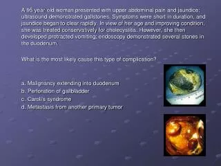
Complications of cholelithiasis • The physical examination might indicate complications of cholelithiasis. • Passage of gallstones from the gallbladder into the common bile duct can result in a complete or partial obstruction of the common bile duct. • Frequently, this manifests as jaundice. • In all races, jaundice is detected most reliably by examination of the sclera in natural for yellow discoloration.
Complications of cholelithiasis • Pancreatitis, another complication of gallstone disease, presents with more diffuse abdominal pain, including pain in the epigastrium and left upper quadrant of the abdomen.
Complications of cholelithiasis • Severe hemorrhagic pancreatitis occurs in 15% patients and carries a high mortality rate because of multisystem organ failure. • In a few patients, the hemorrhagic pancreatic process and retroperitoneal bleeding induce discoloration around the umbilicus (Cullen sign) or the flank (Grey-Turner sign).
Complications of cholelithiasis • Charcot triad • (right upper quadrant pain, fever, and jaundice) • associated with common bile duct obstruction and cholangitis • Additional symptoms: • alterations in mental status and hypotension, indicate Raynaud pentad, a harbinger of worsening, ascending cholangitis.
Causes of cholelithiasis • Prolonged fasting (5-10 days) can result in the formation of biliary sludge (microlithiasis) which resolves by itself when feeding is reestablished - but it can lead to biliary symptoms or gallstone formation
Lab Studies • For patients with uncomplicated cholelithiasis, blood work results usually are normal. • However, labs can detect complications of gallstone disease; complications might alter the course of treatment.
Lab Studies • CBC • chemistry panel, including electrolytes, liver enzymes, and bilirubin. • Choledocholithiasis can manifest with only elevation of serum alkaline phosphatase or bilirubin. • Nearly 50% of patients with symptomatic gallstone disease will have abnormal transaminases
Lab Studies • Serum lipase and amylase levels are helpful in cases of diagnostic uncertainty or suspected concurrent pancreatitis
Imaging Studies • X-rays • Approximately 15% of gallstones are radiopaque and can be visualized on plain x-ray. • A porcelain gallbladder (heavily calcified) should be removed surgically because of increased risk of gallbladder cancer. • Other causes of abdominal pain diagnosed with the assistance of x-rays include perforated viscus, bowel obstruction, calcific pancreatitis, and renal stones.
Imaging Studies • Ultrasound (US) is the most sensitive and specific test for the detection of gallstones. • US provides information about the size of the common bile duct and hepatic duct and the status of liver parenchyma and the pancreas. • Thickening of the gallbladder wall and the presence of pericholecystic fluid are radiographic signs of acute cholecystitis
Imaging Studies • CT scanning often is used in workup of abdominal pain without specific localizing signs or symptoms. • CT scanning is not a first-line study for detection of gallstones because of greater cost and the invasive nature of the test. • When present, gallstones usually are observed on CT scan.
Imaging Studies • HIDA scan does not detect gallstones • HIDA scan identifies an obstructed gallbladder (eg, gallstone impacted in the neck of the gallbladder). • HIDA scan is the most sensitive and specific test for acute cholecystitis. • A poorly contracting gallbladder (biliary dyskinesia) might cause the patient's symptoms, and HIDA scan makes the diagnosis. • Acute acalculous cholecystitis is diagnosed most accurately with HIDA scan.
Treatment • Removal of the gallbladder laparoscopic cholecystectomy is the treatment of choice for symptomatic gallbladder disease • Only gallstones that cause symptoms or complications require treatment
Treatment • There is generally no reason for prophylactic cholecystectomy in an asymptomatic person unless • the gallbladder is calcified • gallstones are > 3cm in diameter
Acute Cholecystitis • Cholecystitis is associated with gallstones in > 90% of cases • Inflammation develops behind a stone impacted in the cystic duct • May be caused by infectious agents (cytomegalovirus, cryptosporidiosis, or microsporidiosis) common in AIDS patients
Acalculous cholecystitis • should be considered in patient with FUO, RUQ pain occurring 2-4 weeks after major surgery
History • Acute attack often follows a large, fatty meal • sudden, steady pain in epigastrium or right hypochondrium - pain may steadily subside over a period of 12-18 hours • vomiting - 75% Of cases • RUQ tenderness associated with muscle guarding and rebound pain
History • Palpable gallbladder 15% of cases • Jaundice 25% of cases • also suggestive of choledocholithiasis • Fever
Labs • WBC - elevated (12-15,000 usuallly) • Total serum bilirubin 1-4mg/dL • Often elevated levels of: • serum aminotransferase • alkaline phosphatase • serum amylase
Imaging Studies • X-ray • may show radiopaque gallstones 15% of cases • HIDA Scan • useful for obstructed cystic duct • reliable if bilirubin < 5mg/dL • Ultrasound • useful for gallstone visulization
Other Conditions • Some disorders that may be confused with acute cholecystitis: • perforated peptic ulcer • acute pancreatitis • appendicitis (high lying appendix) • liver abscess • hepatitis • pneumonia w/pleurisy on right side • myocardial ischemia
The localization of pain and tenderness in the right hypochondrium with radiation to the infrascapular area strongly favors the diagnosis of acute cholecystitis
Treatment • Conservative tx regimen of • TPN • analgesics (Meperidine preferred drug- less spasm of sphincter of Oddi) • antibiotics
Treatment • Due to high rate of recurrence - • cholecystectomy advised • cholecystectomy must be performed when evidence of gangrene or perforation is present
Choledocholithiasis • Choledocholithiasis - common bile duct stones • Occur in 15% of patients with gallstones • Increases with age - in elderly w/gallstones occurrence as high as 50% • Usually condition goes unknown until obstruction occurs
History • History suggestive of biliary colic or jaudice • frequent/recurrent attacks of severe RUQ pain- duration of several hours • severe colic - chills/fever
History • Charcot’s Triad- classic picture of cholangitis • Pain • Fever • Chills
Imaging • The most direct and accurate way to determine the cause, location, and extent of obstruction: • ERCP • percutaneous transhepatic cholangiography
Treatment • Common duct stone in patient with cholelithiasis and cholecystitis is usually treated with endoscopic papillotomy and stone extraction - followed by laparoscopic cholcystectomy
Treatment • Ciprofloxacin, 250mg IV q 12 hours effective tx for cholangitis • alternative tx - mezlocillin, 3g IV q 4 hours with either metronidazole or gentamicin or both • Aminoglycosides should not be used for more than several days due to increased risk of aminoglycoside nephrotoxicity in cholestasis
Primary Sclerosing Cholangitis • Rare disorder • Characterized by diffuse inflammation of the biliary tract leading to fibrosis and strictures of the biliary system • Most common - men aged 20-40
Primary Sclerosing Cholangitis • Associated with histocompatible antigens HLA-B8 and -DR3 or -DR4 - suggestive of genetic etiologic role • Sclerosing cholangitis may occur in AIDs patients from infections caused by CMV, cryptosporidium, or microsporum