Download

1 / 0
0 likes | 184 Views
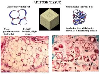
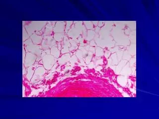
Identification and Characterization of Adipose Tissue M acrophage P opulations in Cats D uring Development of Obesity. Emily C. Graff, DVM, Dipl. ACVP (clinical pathology). U.S. Obesity Trends. Obesity = Adipose Tissue Dysregulation. Adipose tissue- cytology.

E N D