Download
1 / 6
60 likes | 156 Views
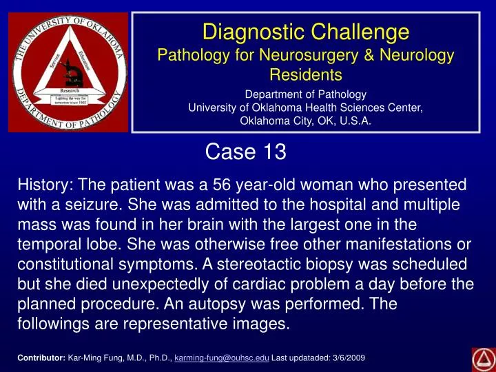
Diagnostic Challenge Pathology for Neurosurgery & Neurology Residents Department of Pathology University of Oklahoma Health Sciences Center, Oklahoma City, OK, U.S.A. Case 13

E N D
Diagnostic ChallengePathology for Neurosurgery & Neurology ResidentsDepartment of PathologyUniversity of Oklahoma Health Sciences Center,Oklahoma City, OK, U.S.A. Case 13 History: The patient was a 56 year-old woman who presented with a seizure. She was admitted to the hospital and multiple mass was found in her brain with the largest one in the temporal lobe. She was otherwise free other manifestations or constitutional symptoms. A stereotactic biopsy was scheduled but she died unexpectedly of cardiac problem a day before the planned procedure. An autopsy was performed. The followings are representative images. Contributor: Kar-Ming Fung, M.D., Ph.D., karming-fung@ouhsc.edu Last updataded: 3/6/2009
Reticulin B C Immunohistochemistry: The large cells are CD20 (+), CD79a (+), and CD3 (-). D
Diagnosis: Diffuse large B-cell type lymphoma. Discussion: The histologic features are highly characteristic for a diffuse large B-cell lymphoma. The features include concentric perivascularinfiltrating large and atypical lymphocytes with expansion of the reticulin network (inset below). The lumen of the blood vessels are indicated by the arrows below. The immunohistochemical profile indicates a B-cell lineage (positive for CD20 and CD79a but negative for CD3).
Multiple lesions (arrows) are present in the gross specimen, a classic finding of primary CNS lymphoma. The hemorrhage in the largest lesion, however, is not always present in lymphoma cases. The patient is not immunocompromised. It should be noted that primary CNS lymphoma can occur in immunocompetent host in the older age group. Those occurring in the immunocomprimised patient often, but not always, occur in younger patients.