Download
1 / 42
560 likes | 1.48k Views
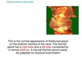
Thyroid, normal, in situ, gross. This is the normal appearance of thethyroid gland on the anterior trachea of the neck. The thyroid gland has a right lobe and a left lobe connected by a narrow i sthmus . A normal thyroid cannot easily be palpated on physical examination.

E N D
Thyroid, normal, in situ, gross This is the normal appearance of thethyroid gland on the anterior trachea of the neck. The thyroid gland has a right lobe and a left lobe connected by a narrow isthmus. A normal thyroid cannot easily be palpated on physical examination.
Thyroid, normal, gross This is a normal adult thyroid gland, which weighs on average from 10 to 30 grams. There is a right lobe and a left lobe, each with an upper pole and a lower pole, connected by an isthmus.
Thyroid, normal, medium power microscopic Normal thyroid seen microscopically consists of follicles lined by a cuboidal epithelium and filled with pink, homogenous colloid. The follicles vary somewhat in size. The interstitium, which may contain "C" cells, is not prominent.
Thyroid, normal, high power microscopic This normal thyroid follicle is lined by a cuboidal follicular epithelium with cells that can add or subtract stored colloid depending upon the degree of stimulation from TSH (thyroid stimulating hormone) released by the anterior pituitary gland. As in all endocrine glands, the interstitium has a rich vascular supply into which hormone is secreted.
Thyroid, normal, "C" cells, immunoperoxidase stain with antibody to calcitonin, medium power microscopic This immunohistochemical stain with antibody to calcitonin identifies the "C" cells (parafollicular cells) of the thyroid interstitium between the follicles, or adjacent to the epithelium of follicles. These parafollicular cells secrete calcitonin, which has minimal impact upon calcium regulation.
Thyroid, atrophy with Hashimoto thyroiditis, gross This symmetrically small thyroid gland demonstrates atrophy. This patient was hypothyroid. This is the end result of Hashimoto thyroiditis. Initially, the thyroid is enlarged and there may be transient hyperthyroidism with Hashimoto thyroiditis, but this is followed by a euthyroid state and then hypothyroidism with eventual atrophy years later from progressive thyroid atrophy. Hashimoto thyroiditis results from abnormal T cell activation and subsequent B cell stimulation to secrete a variety of autoantibodies directed against thyroid.
Thyroid, Hashimoto thyroiditis, low power microscopic This low power microscopic view of a thyroid with an early stage of Hashimoto thyroiditis shows prominent lymphoid follicles. This is an autoimmune disease, and often antithyroglobulin and antimicrosomal (thyroid peroxidase) antibodies can be detected in serum. Other autoimmune diseases such as Addison disease or pernicious anemia may also be present. Both thyroid growth immunoglobulins (TGI) and thyroid stimulating immunoglobulins (TSI) are present, though blocking antibodies to TSI mitigate theireffect.
Thyroid, Hashimoto thyroiditis, high power microscopic This high power microscopic view of thyroid with Hashimoto thyroiditis demonstrates the pink Hürthle cells at the center and right. A lymphoid follicle is at the left. Hashimoto thyroiditis initially leads to painless enlargement of the thyroid, followed by progressive atrophy over following years. Aside from surgery, this is the most common cause for hypothyroidism in adults.
Thyroid, antithyroglobulin antibody, immunofluorescence This is subacute granulomatous thyroiditis (DeQuervain disease), which probably follows a viral infection and leads to a painful enlarged thyroid. This disease is usually self-limited over weeks to months, with transient hyperthyroidism and/or hypothyroidism, and affected patients return to a euthyroid state. Note the presence of large foreign body giant cells with inflammatory destruction of thyroid follicles.
Thyroid, colloid cyst, gross This adult thyroid gland is about normal in size, but there is a larger colloid cyst at the left lower pole and a smaller colloid cyst at the right lower pole. Such cysts could appear as "cold" nodules on a thyroid scintigrafic scan or as an echogenic mass on ultrasound examination. They are incidental benign lesions but can appear as a mass to be distinguished from possible carcinoma.
Thyroid, goiter, patient appearance, gross This patient's anterior neck has an area of pronounced fullness as a consequence of a goiter of the thyroid gland. She is euthyroid, but is bothered by the enlarged thyroid, which presents a cosmetic problem as well as some discomfort.
Thyroid, nodular goiter, gross [CT, NM] This diffusely enlarged thyroid gland above and the one shown below are both somewhat nodular and weigh more than 30 gm. These patients were euthyroid but noted increased fullness to the neck. Multinodular goiter represents the most common cause for an enlarged thyroid gland and the most common disease of the thyroid. Both thyroid lobes are typically diffusely enlarged, but there may be somme asymmetry.
Thyroid, nodular goiter, low power microscopic The follicles are irregularly enlarged, with flattened epithelium, consistent with inactivity, in this microscopic appearance at low power of a multinodular goiter. The earlier phase of a diffuse (non-toxic) goiter leading up to this point may have resulted from either "endemic" goiter (seen in parts of the world where dietary deficiency of iodine may occur) or the uncommon "nonendemic" or sporadic goiter (young adult women are most often affected). Inborn errors of thyroid hormone biosynthesis leading to goiter are extremely uncommon.
Thyroid, Graves disease, low power microscopic [NM] A diffusely enlarged thyroid gland associated with hyperthyroidism is known as Graves disease. At low power microscopically, note the prominent infoldings of the hyperplastic epithelium. In this autoimmune disease the action of thyroid stimulating immunoglobulins (TSI's) predominates over that of thyroid growth immunoglobulins (TGI's). Typical laboratory findings with Graves disease include an increased serum thyroxine but decreased TSH.
Thyroid, Graves disease, high power microscopic Shown at high power, the tall columnar thyroid epithelium with Graves disease lines the hyperplastic infoldings into the colloid. Note the clear vacuoles in the colloid next to the epithelium where the increased activity of the epithelium to produce increased thyroid hormone has led to scalloping out of the colloid in the follicle.
Thyroid, follicular adenoma, gross [XRAY] Here is a surgical excision of a small mass from the thyroid gland that has been cut in half. A rim of slightly darker rim normal surrounding thyroid parenchyma is seen at the left. The mass is well-circumscribed. On physical examination it felt firm. By scintigraphic scan it was "cold." This is a follicular adenoma.
Thyroid, follicular adenoma, gross Here is another follicular neoplasm (a follicular adenoma histologically) that is surrounded by a thin white capsule. It is sometimes difficult to tell a well-differentiated follicular carcinoma from a follicular adenoma. Thus, patients with follicular neoplasms are often treated with subtotal thyroidectomy just to be on the safe side.
Thyroid, follicular adenoma, microscopic Normal, but compressed, thyroid follicles appear at the lower right. The follicular adenoma is at the center to upper left. This adenoma is a well-differentiated neoplasm because it closely resembles normal thyroid tissue. The follicles of the adenoma contain colloid, but there is greater variability in size of the neoplastic follicles than normal. There is no evidence of invasion into the normal thyroid.
Thyroid, follicular carcinoma, microscopic The vascular invasion seen here is evidence for malignancy. The features of papillary carcinoma are lacking, so this is a follicular carcinoma composed of cells that are not highly pleomorphic or hyperchromatic. It can be difficult to tell a follicular carcinoma from an adenoma by histologic appearance alone, and the term "follicular neoplasm" may be utilized. Follicular carcinoma, the second most common thyroid malignancy, tends to be indolent.
Thyroid, papillary carcinoma, gross [CT] Sectioning through a lobe of excised thyroid gland reveals a papillary carcinoma. This neoplasm can be multifocal, as seen here, because of the propensity of this neoplasm to invade lymphatics within thyroid, and lymph node metastases are also common. The larger mass shown here is cystic and contains papillary excresences. These tumors most often arise in middle-aged women. [Image contributed by John Nicholls, MD, Hong Kong University]
Thyroid, papillary carcinoma, low power microscopic [CT] This is the microscopic appearance of a papillary carcinoma of the thyroid. The fronds of tissue have thin fibrovascular cores. The fronds have a papillary pattern. There is no such thing as a papillary adenoma, and all papillary neoplasms of the thyroid should be considered malignant.
Thyroid, papillary carcinoma, high power microscopic This is another papillary carcinoma of thyroid. Note the small psammoma body in the center. The cells of the neoplasm have nuclei with a central clear appearance. Papillary carcinomas are indolent tumors that have a long survival, even when metastases occur. The most favorite site of papillary carcicnoma metastasis is to local lymph nodes in the neck. In fact, some papillary carcinomas may first be detected as a nodal metastasis.
Thyroid, medullary carcinoma, microscopic At the center and to the right is a medullary carcinoma of thyroid. At the far right is pink hyaline material with the appearance of amyloid. These neoplasms are derived from the thyroid "C" cells and, therefore, can have neuroendocrine features such as secretion of calcitonin or other hormones.
Thyroid, medullary carcinoma, amyloid with Congo red stain, microscopic Here the amyloid stroma of the medullary thyroid carcinoma has been stained with Congo red. Medullary carcinomas can be sporadic or familial. The familial kind can be multifocal and are associated with multiple endocrine neoplasia syndrome.
Thyroid, medullary carcinoma, amyloid with Congo red stain, polarized, microscopic This is the Congo red stained amyloid stroma of the medullary carcinoma under polarized light, which produces a pale greenish appearance.
Thyroid, anaplastic carcinoma, gross [CT] The gross appearances of an anaplastic thyroid carcinoma are seen here. This neoplasm is firm and infiltrates surrounding structures, so that externally, on the left, there is no discernible thyroid shape. The cut surface on the right reveals areas of necrosis with cystic change along with the white neoplasm that extends to the margin of resection.
Thyroid, anaplastic carcinoma, medium power microscopic The anaplastic carcinoma shown here is invading into skeletal muscle fibers at the right. This is the most aggressive thyroid cancer, and fortunately the least common form of thyroid cancer.
Thyroid, anaplastic carcinoma, high power microscopic [CT] This anaplastic carcinoma of the thyroid bears no resemblance to normal thyroid tissue--hence the term "anaplastic" to characterize this type of thyroid carcinoma. Note the elongated spindle cells.
Pituitary, normal, gross The normal gross appearance of the pituitary gland removed from the sella turcica is shown here. The larger portion, the anterior pituitary (adenohypophysis), appears in these images toward the top of the gland. The image at the left shows the superior aspect of the pituitary with the stalk coming from the hypothalamus entering it. The inferior aspect of the pituitary is shown at the right. The posterior pituitary (neurohypophysis) is the smaller portion of the gland at the bottom of each image.
Pituitary, normal, low power microscopic The normal microscopic appearance of the pituitary gland is shown here. The adenohypophysis is at the right and the neurohypophysis is at the left.
Pituitary, normal adenohypophysis, medium power microscopic The normal microscopic appearance of the adenohypophysis is shown here. The adenohypophysis contains three major cell types: acidophils, basophils, and chromophobes. The staining is variable, and to properly identify specific hormone secretion, immunohistochemical staining is necessary. A simplistic classification is as follows: The pink acidophils secrete growth hormone (GH) and prolactin (PRL) The dark purple basophils secrete corticotrophin (ACTH), thyroid stimulating hormone (TSH), and gonadotrophins follicle stimulating hormone-luteinizing hormone (FSH and LH) The pale staining chromophobes have few cytoplasmic granules, but may have secretory activity.
Pituitary, normal adenohypophysis, prolactin positive cells, immunoperoxidase stain, medium power microscopic This immunohistochemical stain with antibody to prolactin identifies the specific acidophils in the anterior pituitary that secrete prolactin. Note that they are scattered about amongst other cells of the adenohypophysis.
Pituitary, normal neurohypophysis, medium power microscopic The neurohypophysis shown here resembles neural tissue, with glial cells, nerve fibers, nerve endings, and intra-axonal neurosecretory granules. The hormones vasopressin (antidiuretic hormone, or ADH) and oxytocin made in the hypothalamus (supraoptic and paraventricular nuclei) are transported into the intra-axonal neurosecretory granules where they are released from the neurohypophysis.
Pituitary, anterior lobe, microadenoma, low power microscopic [MRI] This is a microadenoma of the anterior pituitary. Such microadenomas may be present in 1 to 5% of adults. These microadenomas rarely have a significant hormonal output that leads to clinically apparent disease.
Pituitary, anterior lobe, adenoma, high power microscopic [XRAY] Here is a high power microscopic view of an adenohypophyseal adenoma. Endocrine neoplasms are composed of small round cells with small round nuclei and pink to blue cytoplasm. The cells may be arranged in nests or cords, and endocrine tumors also have prominent vascularity as seen here
Pituitary adenoma, gross and microscopic [MRI] The circumscribed mass lesion present here in the sella turcica is a pituitary adenoma. Though pituitary adenomas are benign, they can produce problems either from a mass effect (usually visual problems from pressing on the optic chiasm and/or headaches) or from production of hormones such as prolactin or ACTH. The microscopic appearance of the pituitary adenoma is shown here. Note the monotonous appearance of these small round cells.
Suprasellar craniopharyngioma, gross A large mass extends into the cranial cavity from the region of the sella turcica. This is a suprasellar craniopharyngioma, derived from remnants of Rathke's pouch, that infiltrates into and distorts the surrounding bone. Craniopharyngiomas are much less common than pituitary adenomas. These neoplasms are difficult to eradicate.
Sella turcica, craniopharyngioma, medium and high power microscopic [MRI] A craniopharyngioma is seen here at medium and high power. It is derived from remnants of Rathke's pouch and forms an expanding mass arising in the sellaturcica that erodes bone and infiltrates into surrounding structures. They are difficult to eradicate, even though they are composed of histologically appearing squamoid and columnar epithelium lining cystic spaces filled with oily fluid.
Empty sella syndrome, gross and diagram [MRI] The sella turcica at the base of the skull shown here contains a flattened pituitary at the base, giving the impression of an "empty sella". The diagram indicates how this occurs from herniation of arachnoid (from an arachnoid cyst) into the sella, compressing the pituitary. This may slowly lead to hypopituitarism, if more than 80 or 90% of the adenohypophysis is destroyed. Sometimes, hyperprolactinemia may ensue from a "stalk section" effect.
Lymphocytic hypophysitis, microscopic These medium and high power microscopic views of the anterior pituitary demonstrate mononuclear inflammatory cell infiltrates with loss of acini and interstitial fibrosis. These are features of lymphocytic hypophysitis, a rare autoimmune disorder but a significant cause for hypopituitarism.
END OF LAP ONE DONE BY DR.MOHAMMAD ALHAJ