Download
1 / 2
30 likes | 222 Views
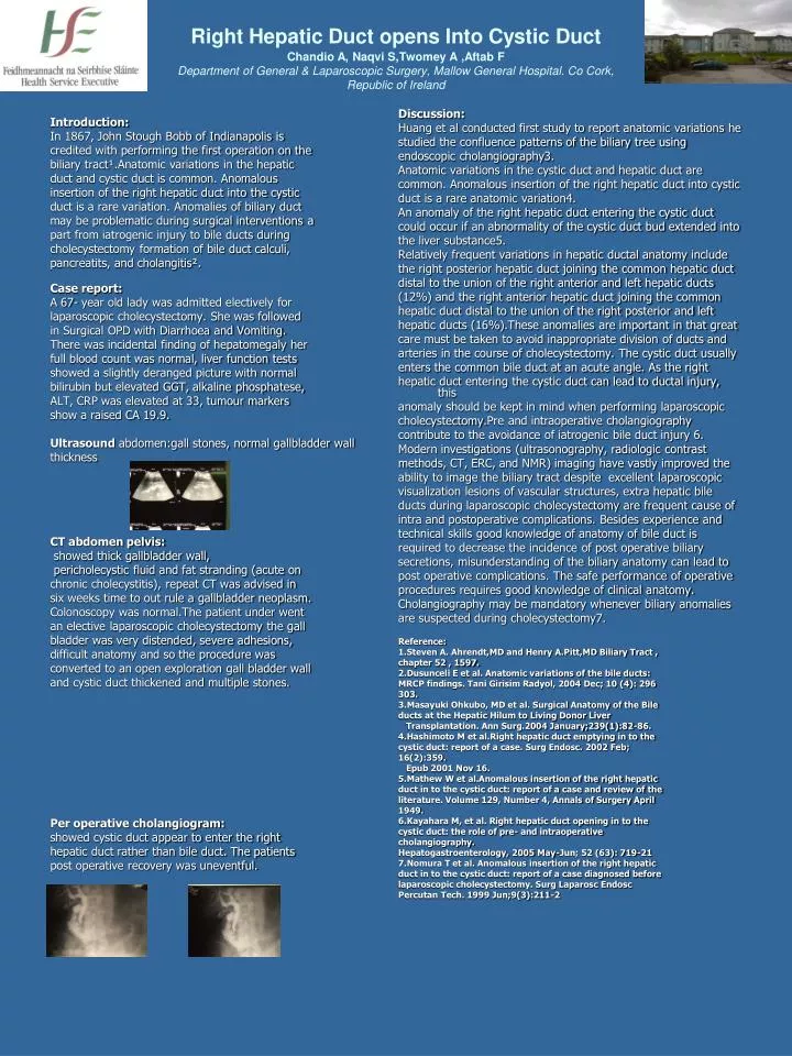
Introduction: In 1867, John Stough Bobb of Indianapolis is credited with performing the first operation on the biliary tract ¹ .Anatomic variations in the hepatic duct and cystic duct is common. Anomalous insertion of the right hepatic duct into the cystic

E N D
Introduction: In 1867, John Stough Bobb of Indianapolis is credited with performing the first operation on the biliary tract¹.Anatomic variations in the hepatic duct and cystic duct is common. Anomalous insertion of the right hepatic duct into the cystic duct is a rare variation. Anomalies of biliary duct may be problematic during surgical interventions a part from iatrogenic injury to bile ducts during cholecystectomy formation of bile duct calculi, pancreatits, and cholangitis². Case report: A 67- year old lady was admitted electively for laparoscopic cholecystectomy. She was followed in Surgical OPD with Diarrhoea and Vomiting. There was incidental finding of hepatomegaly her full blood count was normal, liver function tests showed a slightly deranged picture with normal bilirubin but elevated GGT, alkaline phosphatese, ALT, CRP was elevated at 33, tumour markers show a raised CA 19.9. Ultrasound abdomen:gall stones, normal gallbladder wall thickness CT abdomen pelvis: showed thick gallbladder wall, pericholecystic fluid and fat stranding (acute on chronic cholecystitis), repeat CT was advised in six weeks time to out rule a gallbladder neoplasm. Colonoscopy was normal.The patient under went an elective laparoscopic cholecystectomy the gall bladder was very distended, severe adhesions, difficult anatomy and so the procedure was converted to an open exploration gall bladder wall and cystic duct thickened and multiple stones. Per operative cholangiogram: showed cystic duct appear to enter the right hepatic duct rather than bile duct. The patients post operative recovery was uneventful. Discussion: Huang et al conducted first study to report anatomic variations he studied the confluence patterns of the biliary tree using endoscopic cholangiography3. Anatomic variations in the cystic duct and hepatic duct are common. Anomalous insertion of the right hepatic duct into cystic duct is a rare anatomic variation4. An anomaly of the right hepatic duct entering the cystic duct could occur if an abnormality of the cystic duct bud extended into the liver substance5. Relatively frequent variations in hepatic ductal anatomy include the right posterior hepatic duct joining the common hepatic duct distal to the union of the right anterior and left hepatic ducts (12%) and the right anterior hepatic duct joining the common hepatic duct distal to the union of the right posterior and left hepatic ducts (16%).These anomalies are important in that great care must be taken to avoid inappropriate division of ducts and arteries in the course of cholecystectomy. The cystic duct usually enters the common bile duct at an acute angle. As the right hepatic duct entering the cystic duct can lead to ductal injury, this anomaly should be kept in mind when performing laparoscopic cholecystectomy.Pre and intraoperative cholangiography contribute to the avoidance of iatrogenic bile duct injury 6. Modern investigations (ultrasonography, radiologic contrast methods, CT, ERC, and NMR) imaging have vastly improved the ability to image the biliary tract despite excellent laparoscopic visualization lesions of vascular structures, extra hepatic bile ducts during laparoscopic cholecystectomy are frequent cause of intra and postoperative complications. Besides experience and technical skills good knowledge of anatomy of bile duct is required to decrease the incidence of post operative biliary secretions, misunderstanding of the biliary anatomy can lead to post operative complications. The safe performance of operative procedures requires good knowledge of clinical anatomy. Cholangiography may be mandatory whenever biliary anomalies are suspected during cholecystectomy7. Reference: 1.Steven A. Ahrendt,MD and Henry A.Pitt,MD Biliary Tract , chapter 52 , 1597. 2.Dusunceli E et al. Anatomic variations of the bile ducts: MRCP findings. Tani Girisim Radyol, 2004 Dec; 10 (4): 296 303. 3.Masayuki Ohkubo, MD et al. Surgical Anatomy of the Bile ducts at the Hepatic Hilum to Living Donor Liver Transplantation. Ann Surg.2004 January;239(1):82-86. 4.Hashimoto M et al.Right hepatic duct emptying in to the cystic duct: report of a case. Surg Endosc. 2002 Feb; 16(2):359. Epub 2001 Nov 16. 5.Mathew W et al.Anomalous insertion of the right hepatic duct in to the cystic duct: report of a case and review of the literature. Volume 129, Number 4, Annals of Surgery April 1949. 6.Kayahara M, et al. Right hepatic duct opening in to the cystic duct: the role of pre- and intraoperative cholangiography. Hepatogastroenterology, 2005 May-Jun; 52 (63): 719-21 7.Nomura T et al. Anomalous insertion of the right hepatic duct in to the cystic duct: report of a case diagnosed before laparoscopic cholecystectomy. Surg Laparosc Endosc Percutan Tech. 1999 Jun;9(3):211-2 Right Hepatic Duct opens Into Cystic Duct Chandio A, Naqvi S,Twomey A ,Aftab F Department of General & Laparoscopic Surgery, Mallow General Hospital. Co Cork, Republic of Ireland
CT abdomen pelvis: showed thick gallbladder wall, pericholecystic fluid and fat stranding (acute on chronic cholecystitis), repeat CT was advised in six weeks time to out rule a gallbladder neoplasm. Colonoscopy was normal.The patient under went an elective laparoscopic cholecystectomy the gall bladder was very distended, severe adhesions, difficult anatomy and so the procedure was converted to an open exploration gall bladder wall and cystic duct thickened and multiple stones.