Download
1 / 41
440 likes | 739 Views
Chapter 14: The Digestive System. Functions. Ingestion —taking in food Digestion —breaking food down both physically and chemically Absorption —movement of nutrients into the bloodstream Defecation —rids the body of indigestible waste. Organs.

E N D
Chapter 14: The Digestive System
Functions • Ingestion—taking in food • Digestion—breaking food down both physically and chemically • Absorption—movement of nutrients into the bloodstream • Defecation—rids the body of indigestible waste
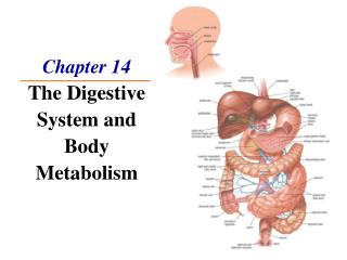
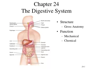
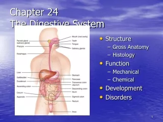
Organs Two main groups: • Alimentary canal (gastrointestinal or GI tract) • continuous coiled hollow tube • Mouth, pharynx, esophagus, stomach, small intestine, large intestine, anus • Accessory digestive organs • Teeth, salivary glands, pancreas, liver, gallbladder
Mouth Anatomy: • Lips (labia) • Cheeks • Hard palate • Soft palate • Uvula • Helps with speech and preventing food/liquid from entering nasal cavity when swallowing
Vestibule • space between lips externally and teeth and gums internally • Oral cavity proper • area contained by the teeth • Tongue • attached at hyoid bone and styloid processes of the skull, and by the lingual frenulum to the floor of the mouth • Tonsils • Palatine & Lingual
Function: • Mastication (= chewing) of food • Digestion by saliva • Swallowing by the tongue • Sense of taste • Gustatory receptors
Pharynx • Only oropharynx and laryngopharynx are part of digestive system • Passageway for air and food • 2 muscle layers alternate contractions to push food down to esophagus (=peristalsis) • Longitudinal inner layer • Circular outer layer
Esophagus • ~10 inches long • Runs from pharynx to stomach through the diaphragm • Passageway for food only • moves food through slow rhythmic squeezing (= peristalsis)
Layers of Alimentary Canal Past the Esophagus 4 layers from inside to outside: • Mucosa • Mostly moist epithelial tissue • Submucosa • Connective tissue with blood vessels, nerve endings, and lymph vessels • Muscularisexterna • Muscle tissue made of inner circular layer and outer longitudinal layer • Serosa • Fluid producing cells make up visceral peritoneum layer that runs into parietal peritoneum (= lining of abdominopelvic cavity)
Nervous System’s Role • Alimentary canal controlled by autonomic nervous system • 2 networks: • Myenteric nerve plexus: between circular and longitudinal muscles • Submucosalnerve plexus: in submucosa layer • Function is to regulate mobility and secretory activity
Stomach • Located on the left side of the abdominal cavity • Food enters at the cardioesophagealsphincter (sphincter = valve) • Food empties into the small intestine at the pyloric sphincter
Regions: • Cardiac region -near the heart • Fundus – rounded part next to the cardiac region • Body - middle • Pylorus - funnel-shaped end
Rugae= internal folds of the mucosa • External regions • Lesser curvature—concave medial surface • Greater curvature—convex lateral surface
Layers of peritoneum attached to the stomach • Lesser omentum- attaches the liver to the lesser curvature • Greater omentum- attaches the greater curvature to the posterior body wall • Sticky wall that collects fat to insulate, cushion, and protect abdominal organs
Physiology • Temporary storage tank for food • Site of food breakdown • The enzyme pepsinogen and hydrochloric acid break down proteins • Gastrin is hormone that stimulates HClsecrection • Alkaline mucus lines the inside of the stomach to protect it from HCl • Delivers chyme (processed food) to the small intestine
Structure of the Stomach Mucosa Figure 14.4c
Small Intestine • Muscular tube extending from the pyloric sphincter to the ileocecal valve • Suspended from the posterior abdominal wall by the mesentery • The body’s major digestive organ • Function: nutrient absorption into the blood
Divisions: • Duodenum • Attached to the stomach • Curves around the head of the pancreas • Jejunum • Middle • Ileum • From jejunum to large intestine
Chemical digestion begins in the small intestine • Enzymes are produced by • Intestinal cells • Pancreas • Pancreatic ducts carry enzymes to the small intestine • Bile, made by the liver, enters through bile duct
3 structural modifications that increase surface area (fom largest to smallest): • Circular folds (plicaecirculares) • Villi – fingerlike projections on circular folds • Microvilli – smaller fingerlike projections on villi that absorb; make the brush border
Large Intestine • Larger in diameter, but shorter in length, than the small intestine • Frames the internal abdomen • Function: absorbs water & processes waste material
Parts: • Cecum- saclike first part of the large intestine • Appendix • Vestigial structure • Accumulation of lymphatic tissue that hangs from the cecum • Colon • Ascending—travels up right side of abdomen • Transverse—travels horizontally • Descending—travels down the left side • Sigmoid—enters the pelvis • Rectum and anal canal • External anal sphincter (skeletal muscle) and internal involuntary sphincter (smooth muscle)
Large Intestine Figure 14.8
No villi present • Goblet cells produce alkaline mucus which lubricates the passage of feces • Muscularisexterna layer is reduced to three bands of muscle called teniae coli • Causes the wall to pucker into pocket like sacs called haustra
Teeth • Function is to masticate (chew) food • Humans have two sets of teeth: • 20 deciduous (baby) teeth by age 2 • 32 permanent teethby age 12
Classification of teeth: • Incisors—cutting • Canines—tearing or piercing • Premolars—grinding • Molars—grinding
Salivary Glands • Three pairs of salivary glands empty secretions into the mouth • Parotid glands • Submandibular glands • Sublingual glands
Saliva • 98% water, 2% electrolytes, mucus, and enzymes • Dissolves chemicals so they can be tasted • Helps to form a food bolus • Your body produces 1-2 liters per day
Pancreas • Located deep to stomach • Produces enzymes that break down carbohydrates • Enzymes secreted into the duodenum • Alkaline fluid mixed with enzymes neutralizes acidic chyme coming from stomach • Produces hormones that regulate blood sugar: • Insulin (takes sugar from blood to tissues) • Glucagon (takes sugar from tissues to blood)
Pancreas Figure 14.1
Liver • Largest gland in the body • Located on the right side of the body under the diaphragm • 4 lobes • Connected to the gallbladder via the common hepatic duct • Functions: • makes bile to break down fat • makes cholesterol to transport fats • makes proteins for blood plasma • detoxifies blood from drugs and alcohol • Stores glycogen, vitamins, and minerals
Liver Figure 14.1
Bile • Yellowish-green fluid produced in the liver • Made of: • Pigments, mostly bilirubin • Made when the liver breaks down old RBCs • Causes yellow coloring of jaundice when there is liver disease • Salts • Cholesterol • Phospholipids • Electrolytes • Function: breaks large fat globules into smaller ones
Gallbladder • Sac found in hollow space of liver • When no digestion is occurring, bile backs up the cystic duct for storage in the gallbladder • When digestion of fatty food is occurring, bile is released into the duodenum from the gallbladder • Gallstones are crystallized cholesterol which can cause blockages